Figure 5. Co-localization of Zbed4 with cell markers in the developing mouse retina. Mouse retinal sections at several developmental
stages were double labeled with antibodies against Zbed4 (green) and different cell markers: A: Upper panel: mouse Brn3a antibody was used to label nuclei of ganglion cells (red). Lower panel: mouse Pax6 antibody was
used to label amacrine, ganglion and Müller cells (red). B: Mouse glutamine synthetase antibody was used to label Müller glial cells (red) at P0, P15, P21, and P30. Acronyms: VZ, ventricular
zone; GCL, ganglion cell layer; OS, outer segments; ONL, outer nuclear layer; INL, inner nuclear layer. Appearance of yellow
in the merged images indicates co-localization of ZBED4 with the different markers. Clearly, Zbed4 is not expressed in ganglion
or amacrine cells of the retina, but is present in Müller cells throughout the life of the mouse.
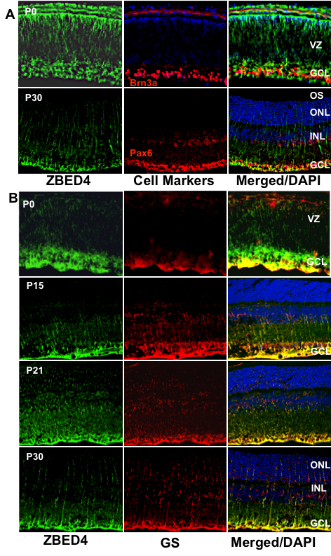
 Figure 5 of
Saghizadeh, Mol Vis 2011; 17:2011-2018.
Figure 5 of
Saghizadeh, Mol Vis 2011; 17:2011-2018.