Figure 3. Developmental pattern of Zbed4 immunoreactivity in mouse retina. From embryonic life through adulthood, Zbed4 is observed
only in Müller cells of the mouse retina. Retinal sections from embryonic day (E)14.5, E16, E18 (A), P0, P5, P8, P11, P15, P21, and P30 (B) animals were incubated with rabbit polyclonal Zbed4 antibody followed by fluorescein isothiocyanate-conjugated goat anti-rabbit
antibodies and 4',6-diamidino-2-phenylindole (DAPI) to visualize nuclei. Merged images of Zbed4 antibody and DAPI stainings
are shown in the right panel of each set. Acronyms: VZ, ventricular zone; GCL, ganglion cell layer; ONL, outer nuclear layer;
INL, inner nuclear layer. Magnification is 400×. Mouse retinal sections of all time points were also reacted with pre-immune
serum followed by secondary antibody as a control for nonspecific binding. Only one of these control sections is shown for
P30. This section was also incubated with a chicken antibody against green/red cone opsin followed by reaction with Alexa
568 goat anti-chicken IgG. In addition, retinal sections from mice of all ages were incubated with Zbed4 antibody that was
pre-absorbed by the peptide used to generate it, and with antibody against green opsin and DAPI. Although the green cones
can be identified by their red color, no staining for Zbed4 was observed, attesting to the specificity of the Zbed4 antibody.
Only one of these control sections is shown for P30.
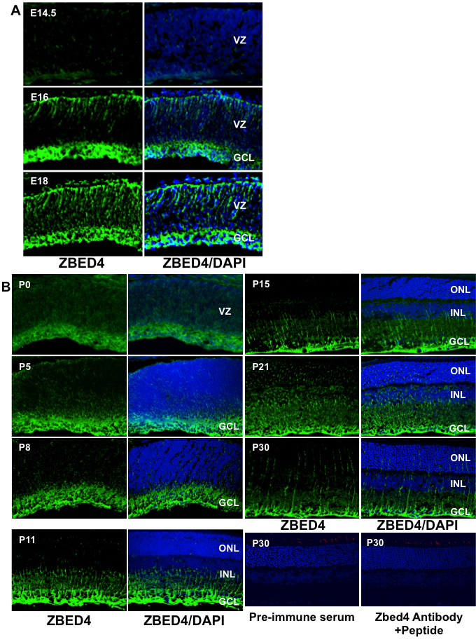
 Figure 3 of
Saghizadeh, Mol Vis 2011; 17:2011-2018.
Figure 3 of
Saghizadeh, Mol Vis 2011; 17:2011-2018.