Figure 1. Effects on proliferation and cell death in human lens epithelial cells (HLECs) exposed to 17β-estradiol at different concentrations
for 24 h. A: The change in number of viable cells (% of control) was determined by the MTT colorimetric assay. B: Difference in the number of mitotic cells (% of control) as evident after staining with Hoechst 33342. C, D: HLECs stained with PI after exposure to 10 µM 17β-estradiol and corresponding control. E: Differences in the number of dead cells (% of control) determined by labeling with PI. Mean±SEM is shown. Experiments were
performed three times in triplicates (n=3) and one representative experimental run for each method is shown. *p<0.05 as compared
to control without 17β-estradiol (0) exposure. Scale bar=100 µm.
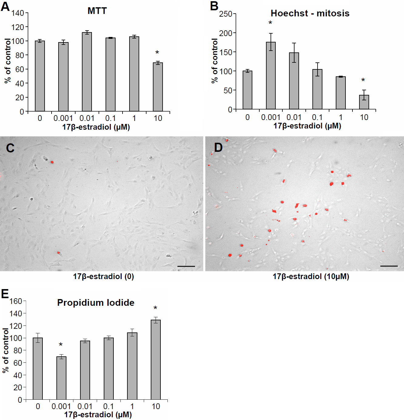
 Figure 1 of
Celojevic, Mol Vis 2011; 17:1987-1996.
Figure 1 of
Celojevic, Mol Vis 2011; 17:1987-1996.