Figure 1. Patient 1 at the age of 8 years, after a bilateral medial rectus recession at the age of 2.5 years. A-F show the eye position at different attempted movements. The eye position at upgaze is shown in A. B depicts the eye position at right gaze. C shows the eyes in primary position. The eye position in left gaze, downgaze and under the condition of convergence is demonstrated
in D, E, F, respectively.
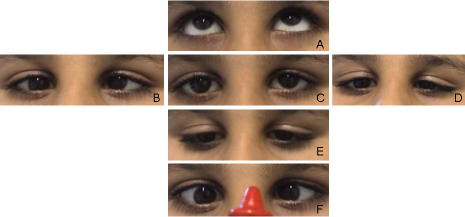
 Figure 1 of
Volk, Mol Vis 2011; 17:1978-1986.
Figure 1 of
Volk, Mol Vis 2011; 17:1978-1986.