Figure 1. Clinical and investigation pictures of patient 1. A: Color fundus photograph of right eye showing scarred choroidal neovascularization (CNV). B: Pretreatment color fundus photograph of the left eye showing retinal crystals with active CNV (arrow) and sub retinal hemorrhage.
C: Fundus fluorescein angiography (FFA) of right eye shows staining. D: FFA of the left eye shows areas of window defects corresponding to areas of RPE and choriocapillaris atrophy with blocked
fluorescence along the areas of crystalline deposits and subretinal hemorrhage. Hyperfluorescence spot (leaked fluorescein)
is seen on right eye subfoveal region, suggestive of active CNV. E: SD-OCT of right eye showed foveal contour with scarred subfoveal CNV. F: SD-OCT of left eye showed loss of foveal contour with increased retinal thickness with an active sub foveal CNV (arrow).
G, H: Multifocal ERG revealed grossly reduced central and paracentral ring responses with reduced peripheral ring response in
both eyes. I, J: Visual field defects were also noted on Humphrey visual field perimetry. K, M: Color fundus photo and OCT of right eye at last follow-up. L: Post Ranibizumab treated color fundus photograph of left eye showed scarred CNV as confirmed by OCT (N) at last follow-up.
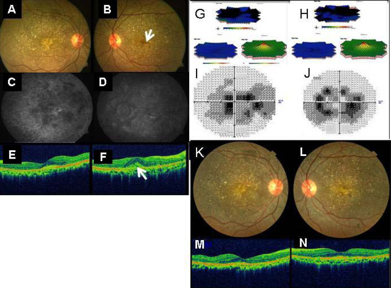
 Figure 1 of
Mamatha, Mol Vis 2011; 17:1970-1977.
Figure 1 of
Mamatha, Mol Vis 2011; 17:1970-1977.