Figure 5. Photographs of the nails and
radiographs of knees, hips and elbows of some of the subjects. Nail
involvement in these cases includes dystrophic, thin, hypoplastic
discolored nails with longitudinal ridges. Case III-2 shows decreased
creases over the skin of distal interphalangeal joints, which is a
sensitive sign of digital involvement in this patient (A).
Thumbs are most severly affected in case III-7 (B), and IV-9 (C).
The
radiographs of knee involvement in Case III-2 shows bilateral
involvement of the patella, with hypoplastic, higher than normal
missplaced patella (D-F). A small 6 mm bone fragment can
be observed on the lateral border of right patella (E). Knee
radiographs of Case III-7 show slight bilateral patellar hypoplasia,
mostly on the transverse diameter (G and H). Hip
radiography of Case IV-2 shows loss of the normal concavity at the
junction between the head and femoral neck bilaterally. The radiographs
reveal an elbow involvement of subject IV-2 (J and K)
and include a prominent medial epicondyle and hypoplasia of the
capitellum on both sides (J and K). They also show a
hypoplasia of the lateral epicondyle and capitellum with a slight
deformity of the radial head (J and K). Underdeveloped
triceps and prominent medial epicondyle are observed (L).
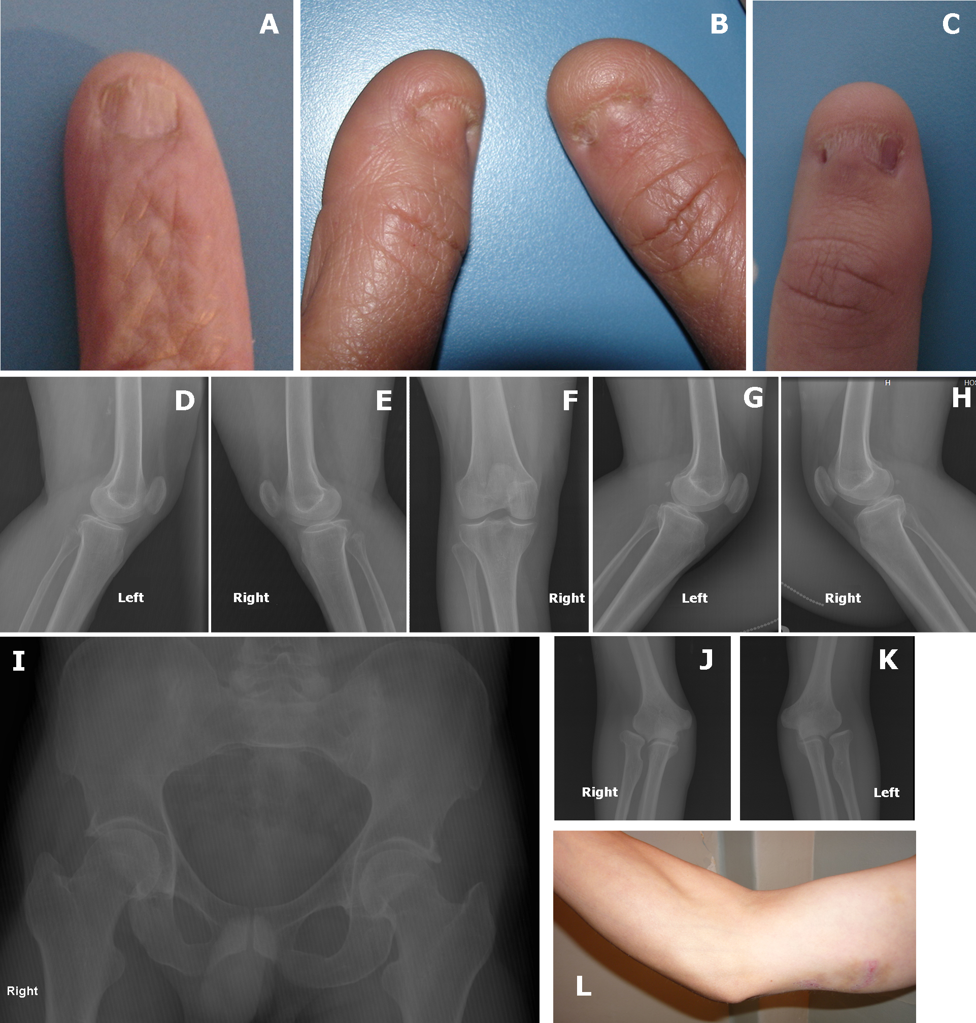
 Figure 5 of Romero, Mol Vis 2011; 17:1929-1939.