Figure 2. Photographs of the optic discs
of glaucomatous eyes and visual fields of the proband. The optic disc
photograph shows that the neuroretinal rim was lost slightly in her
inferior temporal optic disc sector. The left eye shows greater loss of
neuroretinal rim (B, arrow) than the right eye (A). The
visual field shows a nasal step in the right eye (C) and a
superior arcuate scotoma in the left eye (D).
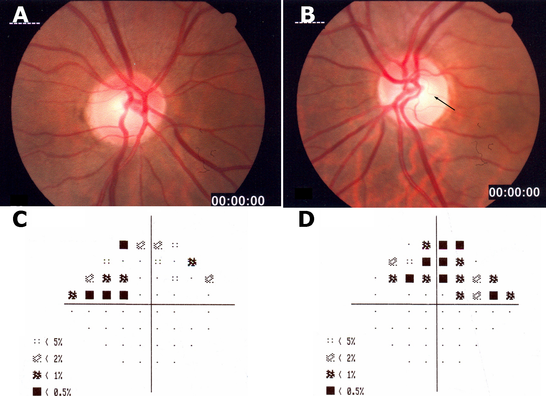
 Figure 2 of Romero, Mol Vis 2011; 17:1929-1939.