Figure 4. The shape changed after LPS
stimulation. MyD88 and NF-κB were expressed differently before and
after LPS stimulation of C3H/HeN mouse peritoneal macrophages. A:
Three
hours after LPS stimulation, the cell elongated significantly. B:
Six
hours after LPS stimulation, the cell showed extended pseudopodia
and protrusions. C: Twelve hours after LPS stimulation, the
cell extended a large number of pseudopodia and protrusions and the
size of cell reached its peak. D: In unstimulated C3H/HeN mouse
peritoneal macrophages without LPS stimulation, MyD88 were expressed in
the cytoplasm and nuclei. E: In C3H/HeN mouse peritoneal
macrophages without LPS stimulation, NF-κB was expressed in the
cytoplasm. F: One hour after LPS stimulation of C3H/HeN mouse
peritoneal macrophages, NF-κB were expressed in the nuclei. G:
NF-κB was expressed in the cytoplasm after the blockage of TLR4 with
MTS510.
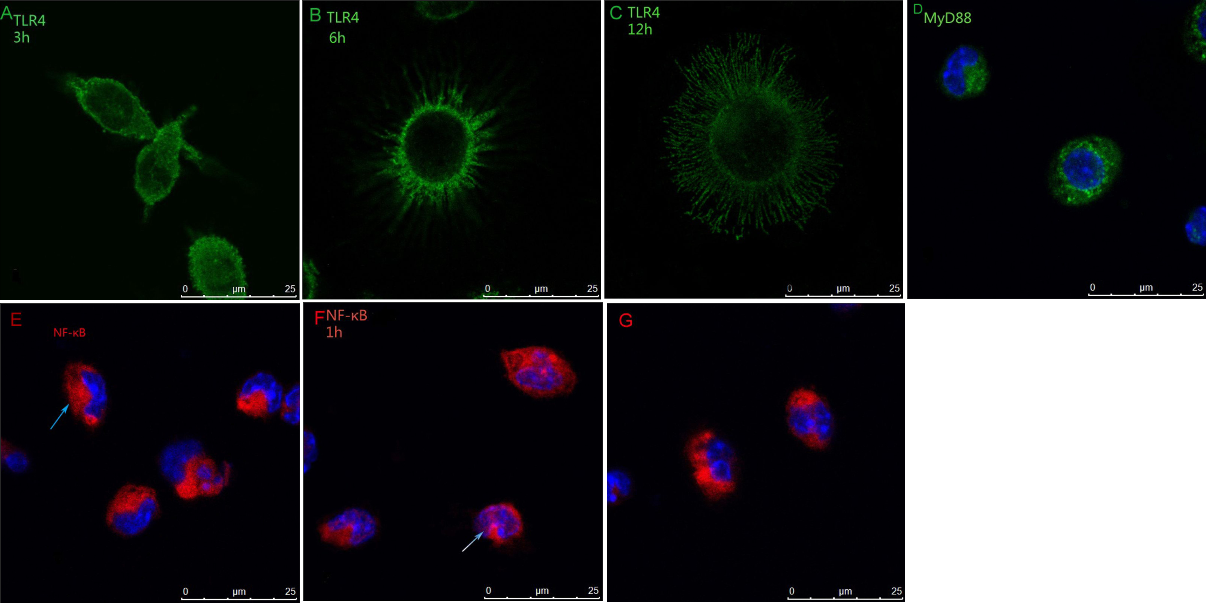
 Figure 4 of Wang, Mol Vis 2011; 17:170-176.