Figure 7. Histological profiles of retinal, hepatic, and renal tissue sections at 90 days. Retinal, hepatic, and renal tissue sections
were normal, and no inflammatory cell infiltration was observed (A-C). Immunohistochemical studies indicated that CD38+ cells were not detected in the retinal, hepatic, and renal tissue sections
(E-G), respectively. In positive controls, the expression level of CD38 rised significantly. CD38+ cells (arrowheads) were detected
in the hepatic tissue sections (D and H), respectively.
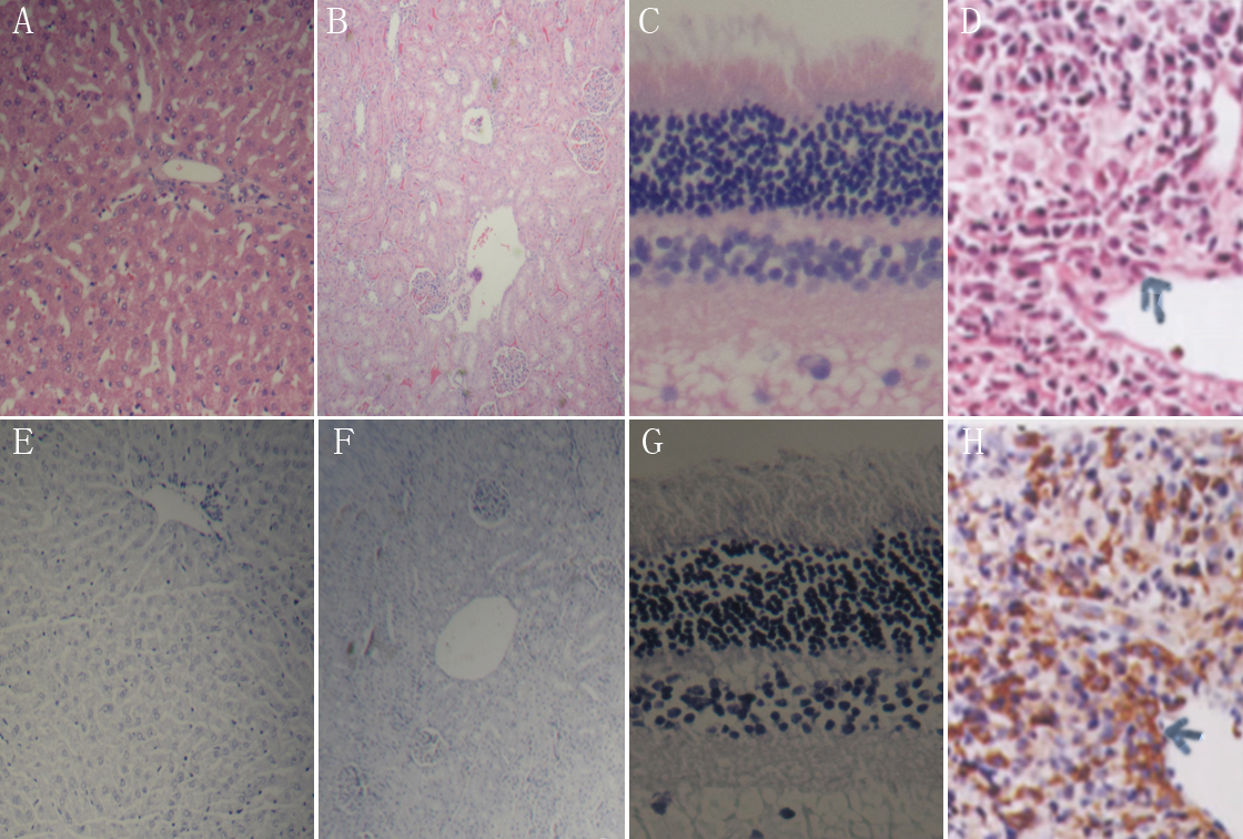
 Figure 7 of
Ge, Mol Vis 2011; 17:1918-1928.
Figure 7 of
Ge, Mol Vis 2011; 17:1918-1928.