Figure 6. Histologic profiles of the
corneal buttons . Corneal buttons were collected on day 14 (A-E).
Corneal
button sections of normal (A),control (B), Vector
(C), endostatin (D), and RGDRGD-endostatin (E)
groups
were stained with hematoxylin and eosin. Expression of VEGF in
the corneal buttons was detected by immunohistochemistry (F-J).
High
levels of VEGF were detected in the control and vector groups,
respectively (G-H); by contrast, only minute levels were
detected in the endostatin and RGDRGD-endostatin gene groups (I-J),
respectively
. Light micrographs of CD31-stained corneas in an
untreated eye after corneal denudation and the effects of
RGDRGD-endostatin gene delivery on experimental corneal angiogenesis on
day 14. Representative light micrographs of CD31-stained corneas given
normal, control,vector,endostatin,or the RGDRGD-endostatin expression
cassette are shown (each group; n=3; K-O).The absolute
normal control group showed no neovascularization, the experimental
normal and vector groups showed neovascular formation (arrowheads), the
endostatin group showed less neovascular formation,while the
RGDRGD-endostatin group showed significantly less vessels and smaller
areas of neovascularization compared with endostatin-treated corneas.
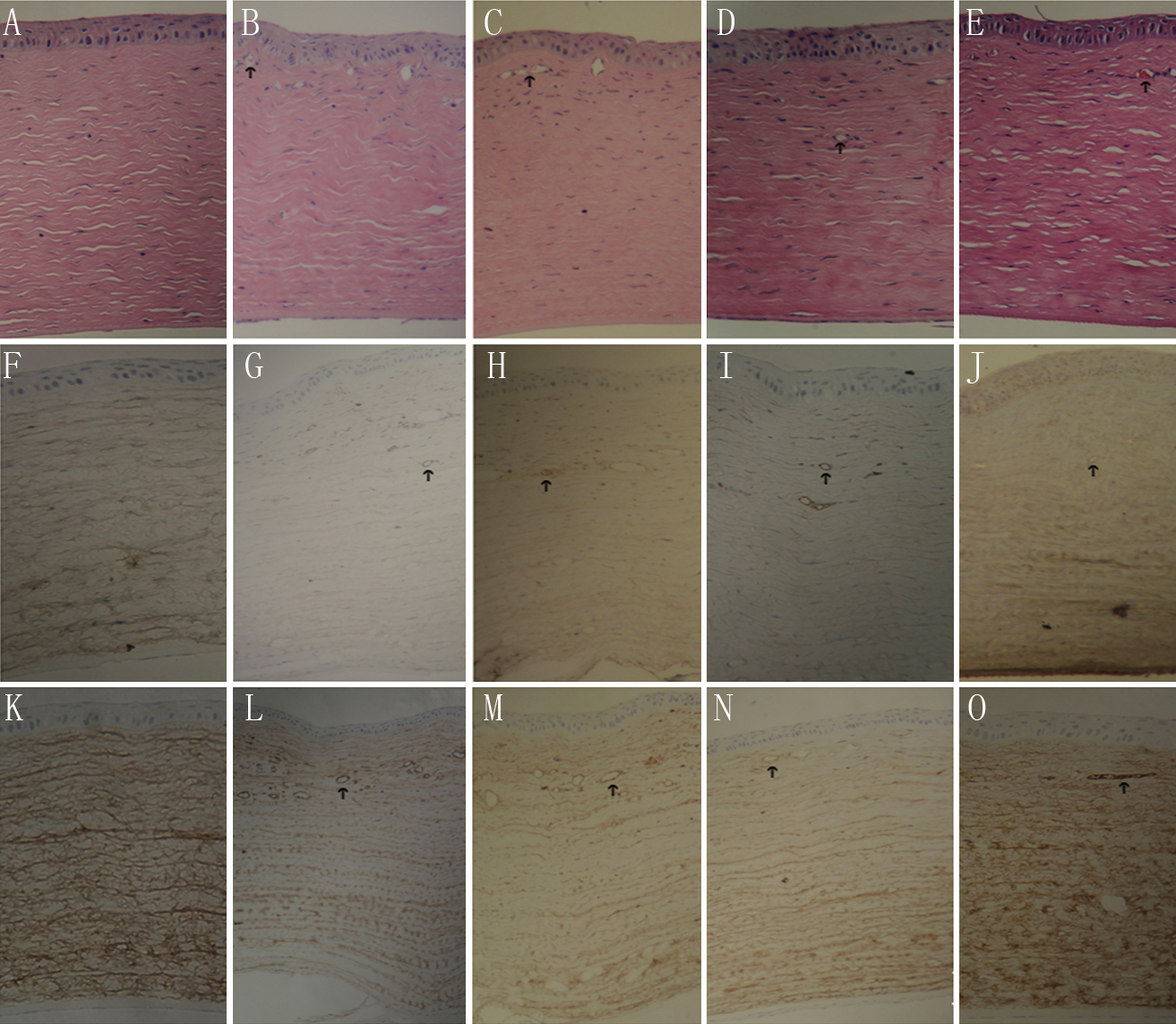
 Figure 6 of Ge, Mol Vis 2011; 17:1918-1928.