Figure 1. Schematic diagram of corneal
transplantation procedure. A: The anterior lamella was
dissected from the rabbit cornea. B: The prepared fresh cat
corneal lamella/ACS were sewn into place for TLKP. C: The graft
bed for PKP was prepared in the central cornea. D: A
full-thickness donor cat cornea was placed in the recipient bed.
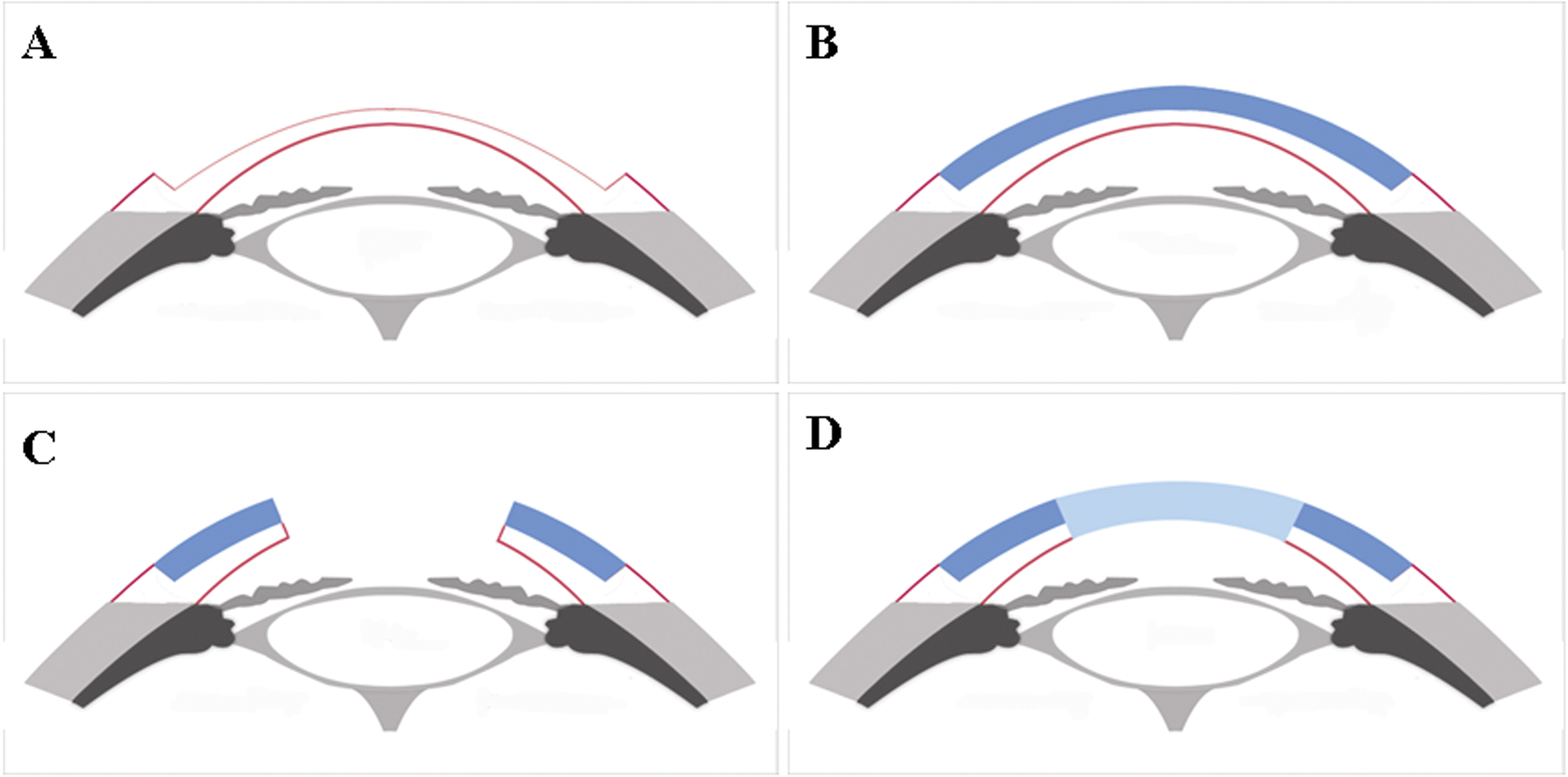
 Figure 1 of Li, Mol Vis 2011; 17:1909-1917.