Figure 5. Glial fibrillary acidic protein
(GFAP) localization in retinal flat-mounts of alkali injured mice at 14
days post-injury. A: Prominent GFAP expression as observed in
this image montage is visible throughout the retina most notably
upregulated in astrocytes. B: Enlarged image of activated
Muller cells. C: Representative enlarged image of astrocytes
showing GFAP staining. For orientation purposes, the optic nerve is
marked with a white semi-circular line. The scale bar in the montage
image (A) is 215 μm. The scale bars in the enlarged images are
70 μm.
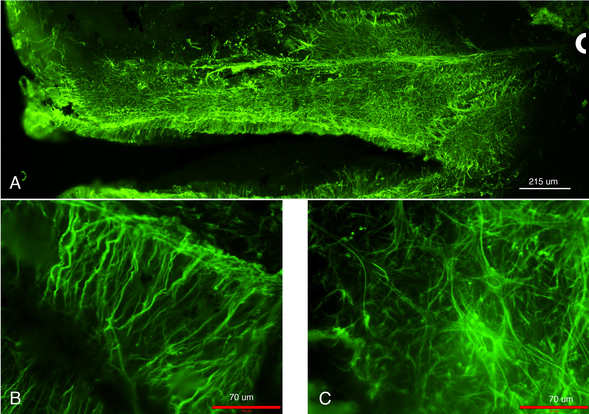
 Figure 5 of Paranthan, Mol Vis 2011; 17:1901-1908.