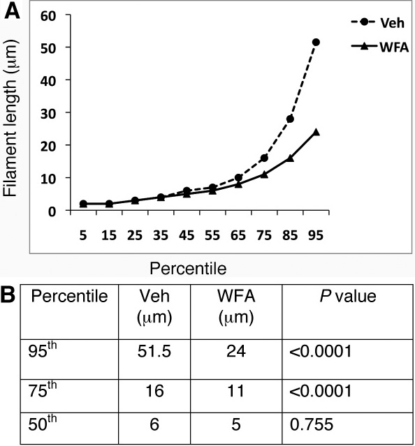
Figure 4. Quantitative assessment of glial fibrillary acidic protein (GFAP) filaments. Mice were subjected to corneal alkali injury
with limbal and corneal epithelial cell debridement and treated daily with dimethyl sulfoxide (DMSO, Veh) or 2 mg/kg withaferin
A (WFA) for 14 days. Tissue sections of injured eyes from vehicle-treated and WFA-treated mice (n=15/group) were stained with
antibodies to GFAP and filaments quantified as previously reported [
21]. Graphical representations of GFAP filament lengths as percentiles are shown between vehicle-treated and WFA-treated samples
(
A). Significant differences were noted above the 50th percentiles between these two groups (
B).
 Figure 4 of
Paranthan, Mol Vis 2011; 17:1901-1908.
Figure 4 of
Paranthan, Mol Vis 2011; 17:1901-1908.