Figure 3. Fourteen days post-injury
gliosis in the retina. Mice were subjected to corneal alkali injury
with limbal and corneal epithelial cell debridement and treated daily
with dimethyl sulfoxide (DMSO, Veh) or 2 mg/kg withaferin A (WFA) for
14 days. A-C: Tissue sections of eyes from uninjured (Cont),
injured (veh) and WFA treated mice were stained with antibodies to
glial fibrillary acidic protein (GFAP; green). Nuclei were stained with
4′, 6-diamidino-2-phenylindole (DAPI, blue) and fluorescent staining
was visualized with a Nikon TE2000 microscope. D: western blot
analysis of GFAP expression in soluble extracts of retina samples from
uninjured (cont), injured (Veh) and WFA treated mice. Abbreviations:
GCL represents ganglion cell layer; INL represents inner nuclear layer;
ONL represents outer nuclear layer.
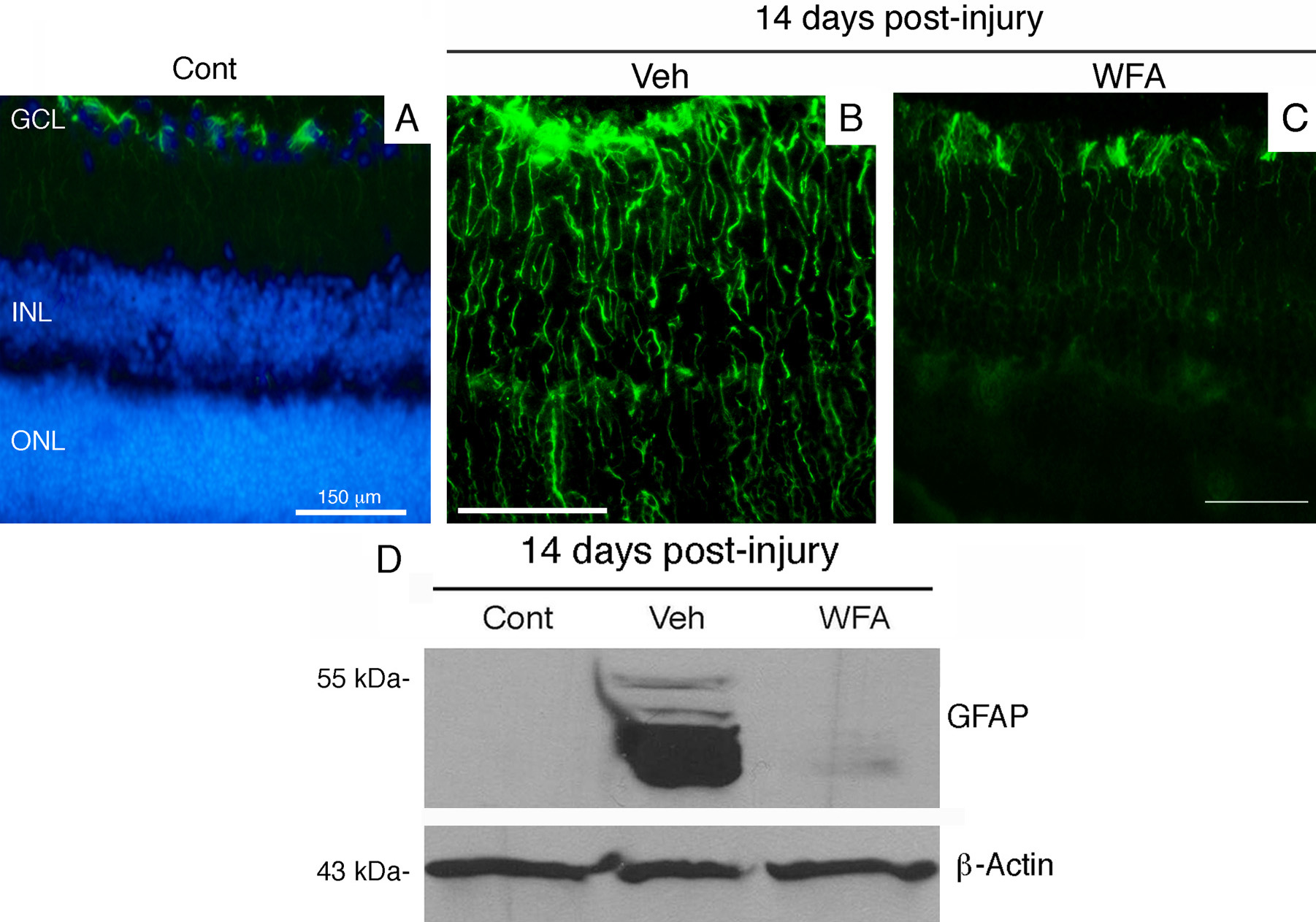
 Figure 3 of Paranthan, Mol Vis 2011; 17:1901-1908.