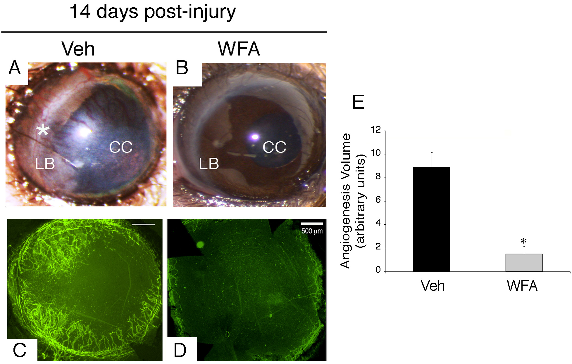
Figure 2. Neovascularization in the cornea
at 14 days post-injury. Mice were subjected to corneal alkali injury
with limbal and corneal epithelial cell debridement and treated daily
with dimethyl sulfoxide (DMSO, Veh) or 2 mg/kg withaferin A (WFA) for
14 days.
A-B: Representative images of anterior segment showing
phenotypic representation of corneal neovascularization with and
without drug treatment. An extensive network of new blood vessels
extends from the limbus (LB, asterisk) into the central cornea (CC) in
Veh sample.
C-D: Whole mount staining of corneas labeled with
anti-CD31-fluorescent antibody (green) show an extensive network of new
blood vessels in vehicle (Veh) sample with potent inhibition in sample
treated with WFA (
D).
E: Quantification of
neovascularization from each group of mice (n=14) was performed as
previously described [
20].
Error
bars represent standard deviation (SD); * p<0.05 by the
Mann–Whitney U test.
 Figure 2 of Paranthan, Mol Vis 2011; 17:1901-1908.