Figure 1. Mouse alkali burn injury model
for corneal neovascularization and retinal gliosis. Corneal alkali
injury with limbal and corneal epithelial cell debridement induces two
distinct ocular pathologies that are robustly elaborated at 14 days
post injury: 1) gliosis in the retina (left panel) and 2)
neovascularization in the cornea (right panel). A: Reactive
Müller cells labeled with glial fibrillari acidic protein (GFAP)
antibody (green) in injured sample from thin tissue section. Cell
nuclei were stained with 4′, 6-diamidino-2-phenylindole (DAPI, blue). B:
Representative
image of a segment from whole-mount injured cornea
labeled with CD-31+ antibody (green) showing
neovascularization from pre-existing limbal vessels (arrowheads). The
cornea image was taken at 10× magnification and the retina image at
30×. Abbreviations: GCL represents glial cell layer; INL represents
inner nuclear layer; ONL represents outer nuclear layer; Epi represents
epithelium.
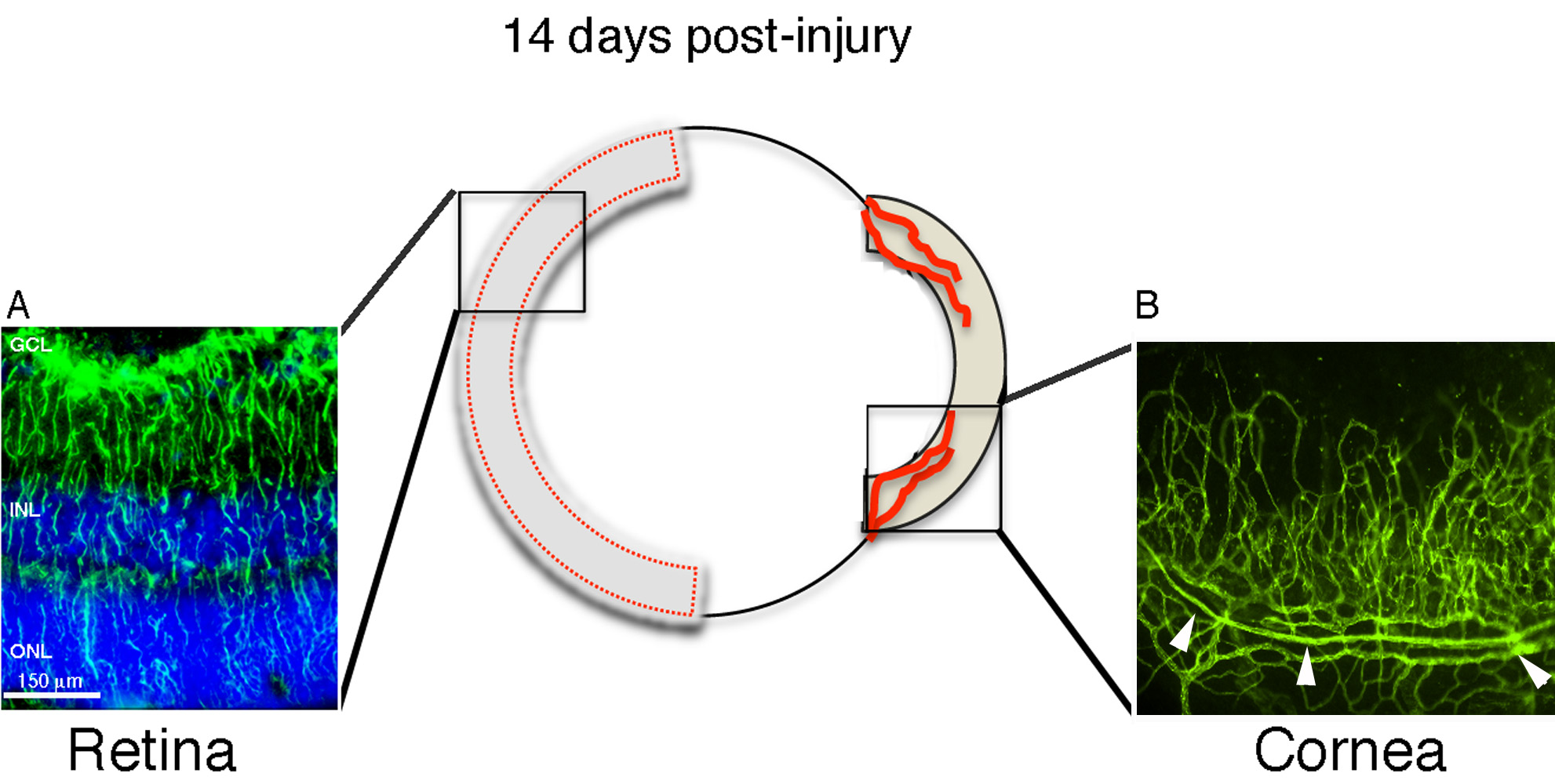
 Figure 1 of Paranthan, Mol Vis 2011; 17:1901-1908.