Figure 3. Comparison of an autosomal
recessive spastic ataxia of Charlevoix-Saguenay patient and a healthy
subject. The patient (left) shows an increase of retinal nerve fiber
layer thickness in the optical coherence tomograph (A), and
moderately increased visibility of the retinal nerve fiber layer in the
stereophotograph (B) of the optic disc and in the monochromatic
red-free digital fundus photograph (C), compared with healthy
eye (D-F).
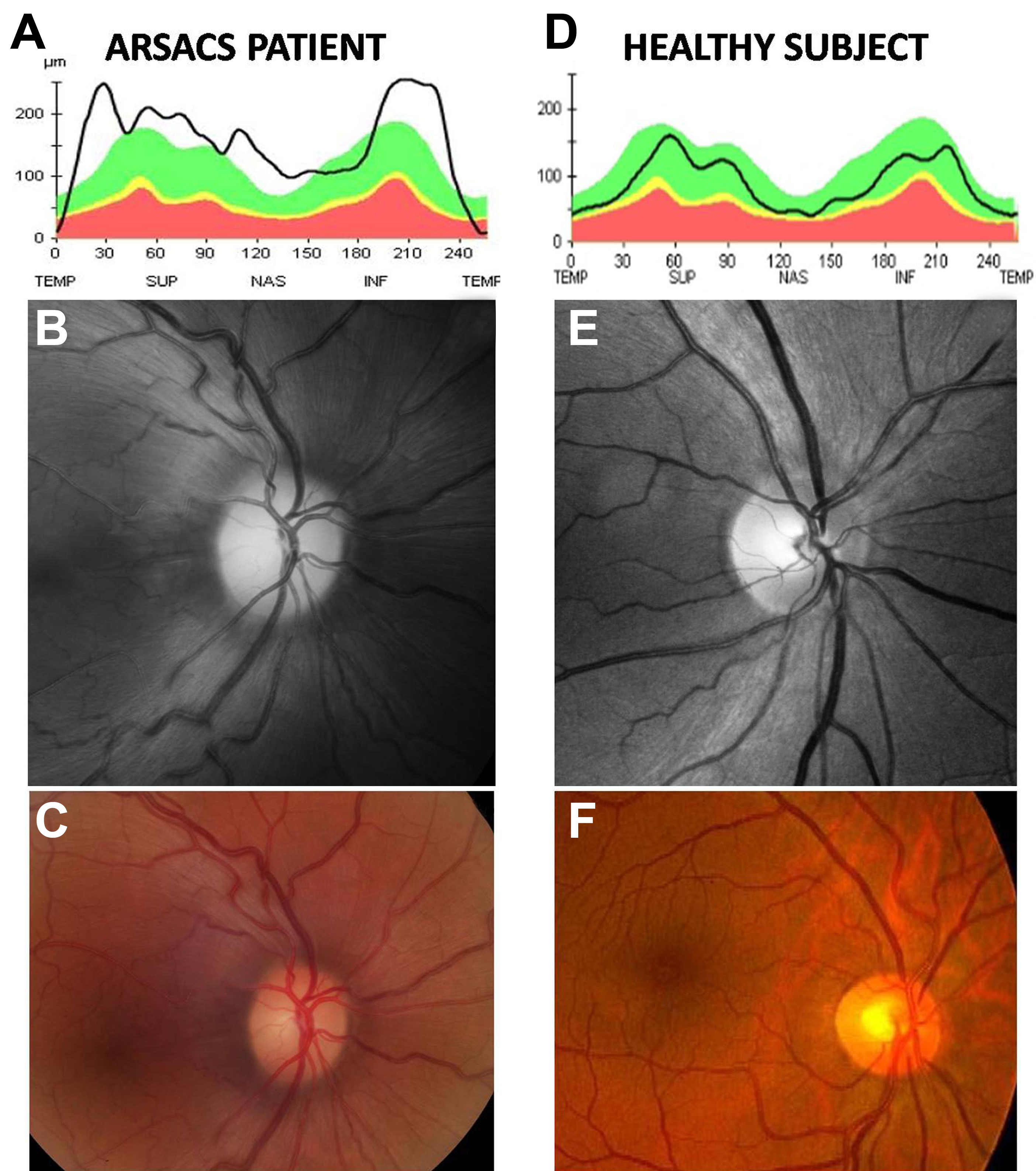
 Figure 3 of Pablo, Mol Vis 2011; 17:1871-1876.