Figure 2. Monochromatic and stereophotographs in autosomal recessive spastic ataxia of Charlevoix-Saguenay 198 patient. A: A monochromatic photograph of retinal nerve fiber layer in patient 4 (left eye) shows increased visibility of fibers and
a thin sector defect (included between arrows). B: A stereophotograph of the same eye shows the telltale yellow discoloration of the retinal nerve fiber layer streaks.
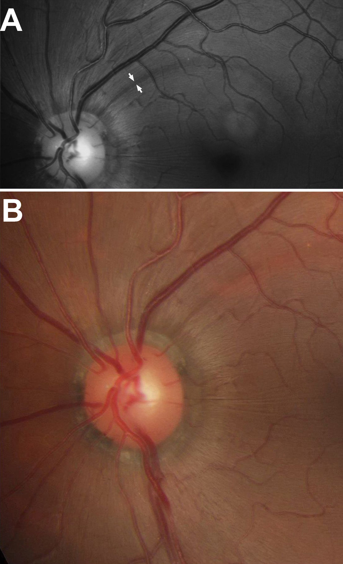
 Figure 2 of
Pablo, Mol Vis 2011; 17:1871-1876.
Figure 2 of
Pablo, Mol Vis 2011; 17:1871-1876.