Figure 2. Immunolocalization of high-mobility group box −1 (HMGB1) in control (upper row of panels) and diabetic (lower row of panels)
retinas. Immunofluorescence using isolectin B4 as a vascular marker (red), a nuclear marker 4’,6-diamidino 2-phenylindole
(DAPI), and HMGB1 antibody showed increased expression of HMGB1 in diabetic retina in comparison to the control. HMGB1 is
localized mainly in ganglion cells (GC), inner nuclear layer (INL), and retinal vasculatures (arrows). Abbreviations: IPL
represents inner plexiform layer; OPL represents outer plexiform layer; ONL represents outer nuclear layer.
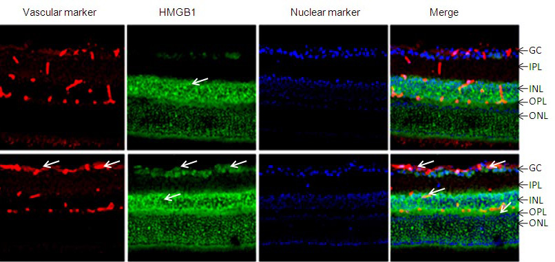
 Figure 2 of
El-Asrar, Mol Vis 2011; 17:1829-1838.
Figure 2 of
El-Asrar, Mol Vis 2011; 17:1829-1838.