Figure 4 of
Raju, Mol Vis 2011; 17:7-15.
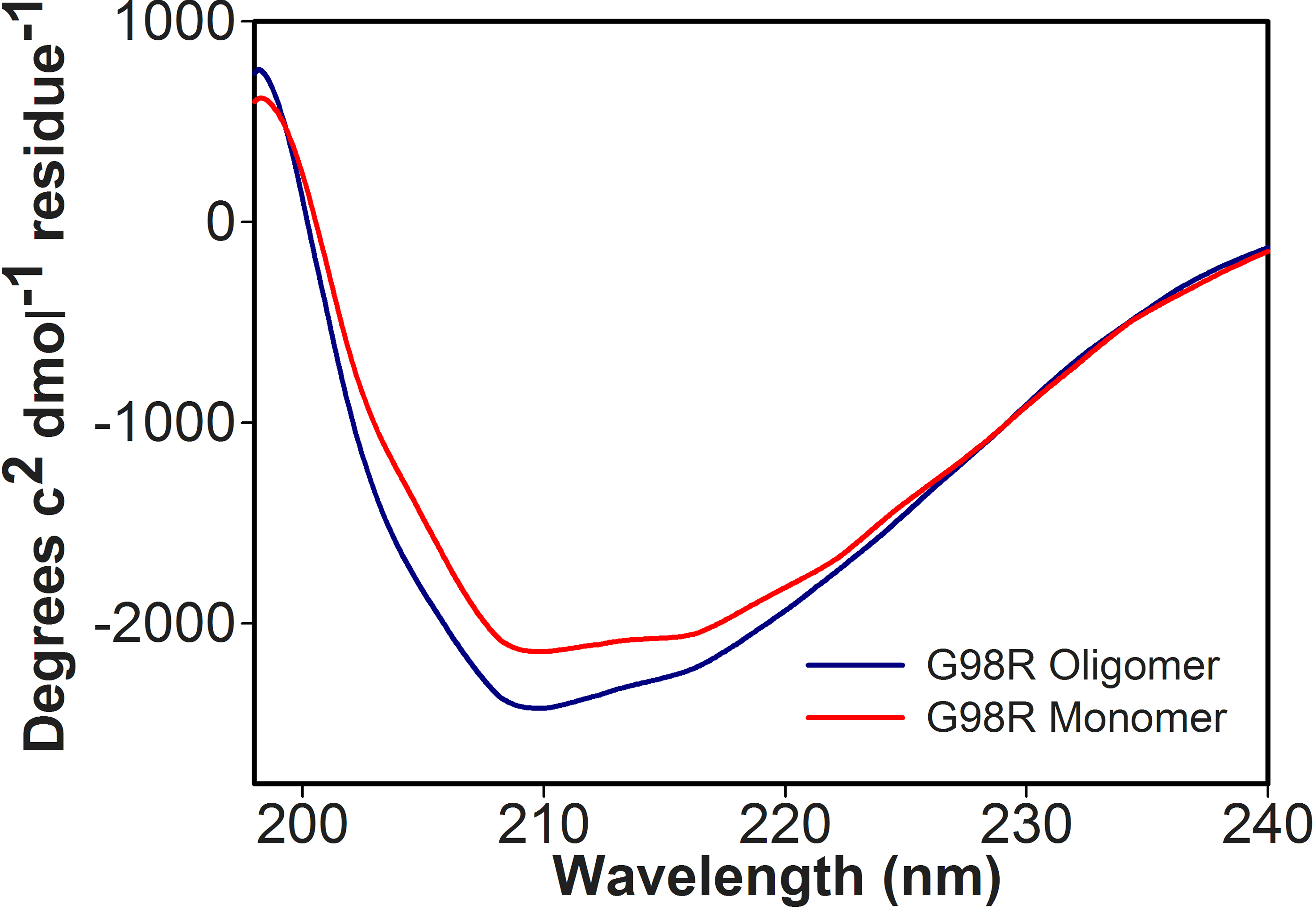
Figure 4.
Far-UV Circular dichroism (CD) spectra of αAG98R-crystallin monomeric protein. CD spectra were recorded using 0.1 mg/ml protein in a 0.2 cm cell path length cuvette at 25 °C. The spectra shown represent an average of six scans.
 Figure 4 of Raju, Mol Vis 2011; 17:7-15.