Figure 2. Size exclusion chromatography
profile and immunoblot showing monomers of αAG98R-crystallin. A:
Size-exclusion
chromatography of three concentrations of
αAG98R-crystallin. αAG98R-crystallin was injected to a TSK-5000 PWXL
column (7.6 mm×30 cm) in 3 concentrations and the elution profile was
recorded by following the 280 nm absorbance. Blue, 0.5 mg/ml; Black,
0.2 mg/ml and Red, 0.1 mg/ml. B: SDS–PAGE and immunoblot of
dissociated subunits of αAG98R-crystallin protein obtained during size
exclusion chromatography analysis. Lane1, Marker proteins; lane 2,
αAG98R-crystallin stained with Coomassie Blue; lane 3, western blot of
lane 2 sample probed with anti-αA-crystallin.
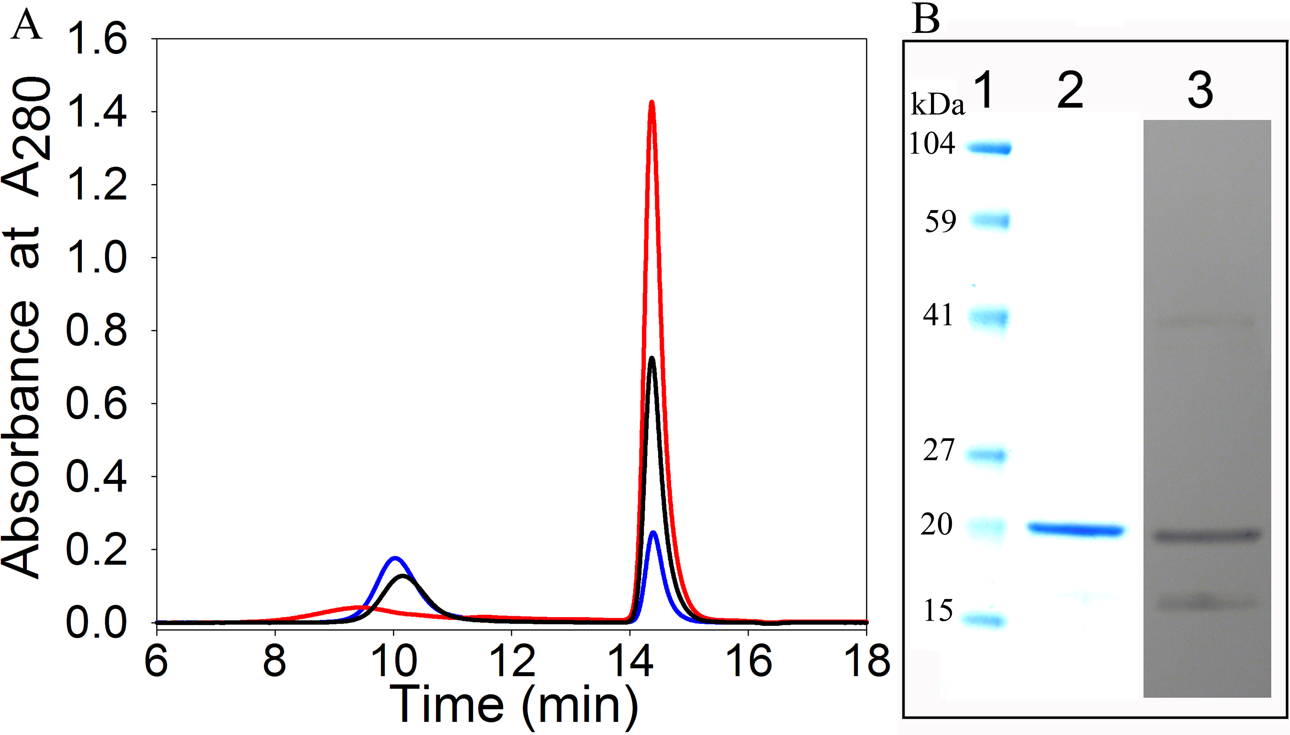
 Figure 2 of Raju, Mol Vis 2011; 17:7-15.