Figure 1. Electrophoretic and size
exclusion chromatography profile of wild-type and αAG98R-crystallin. A:
Comassie
Blue–stained SDS–PAGE of G98R mutant and wild-type
αA-crystallin, showing >98% purity of the crystallins used in the
study. Lane 1, protein markers; Lane 2, wild-type αA-crystallin; and
Lane 3 αAG98R-crystallin. B: Size exclusion chromatography
profile of αAG98R-crystallin and wild-type αA-crystallin. 100 μg of
protein at 1mg/ml concentration in phosphate buffer was injected into a
TSK-G5000PWXL gel filtration column (7.6 mm×30 cm).
Fractions of 0.75 ml were collected. The mutant G98R protein (red)
shows two peaks, one at 9.5 min, corresponding to the oligomer, and
another peak at 15 min, corresponding to monomer mass. Wild-type
α-crystallin (blue) did not show two peaks.
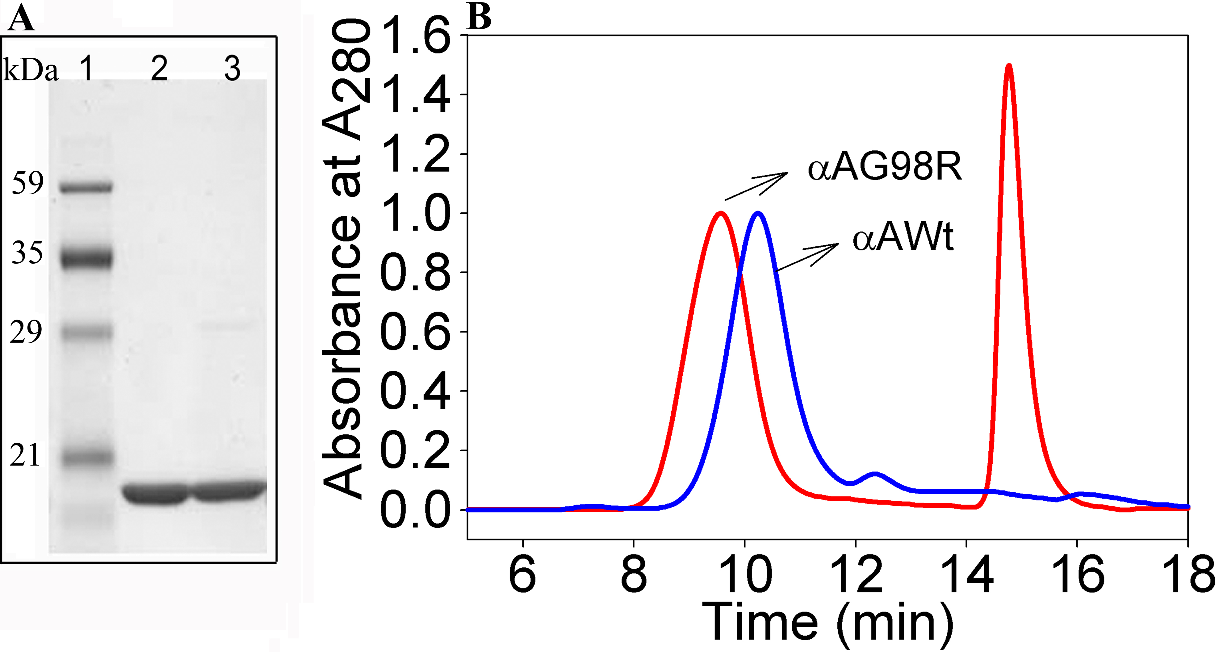
 Figure 1 of Raju, Mol Vis 2011; 17:7-15.