Figure 2. Differential expression of
mitochondrial proteins in early experimental autoimmune uveitis retina.
On day 7 after immunization, mitochondria were isolated from retinas of
B10.RIII mice induced with experimental autoimmune uveitis (EAU) and
from control animals. The EAU and control samples were then
differentially labeled with cyanine dyes (Cy3 and Cy5) and resolved
using a single 2D gel. An internal standard containing equal amounts of
each mitochondrial sample labeled with Cy2 was also used. The two
images from both samples were overlaid, and the gel was scanned using a
highly sensitive typhoon imager and processed by analysis software. The
numbered spots were the differentially regulated proteins in EAU
samples and these spots were picked for Mass Spectrometry analysis. The
differentially expressed proteins were identified by MALDI-TOF/MS.
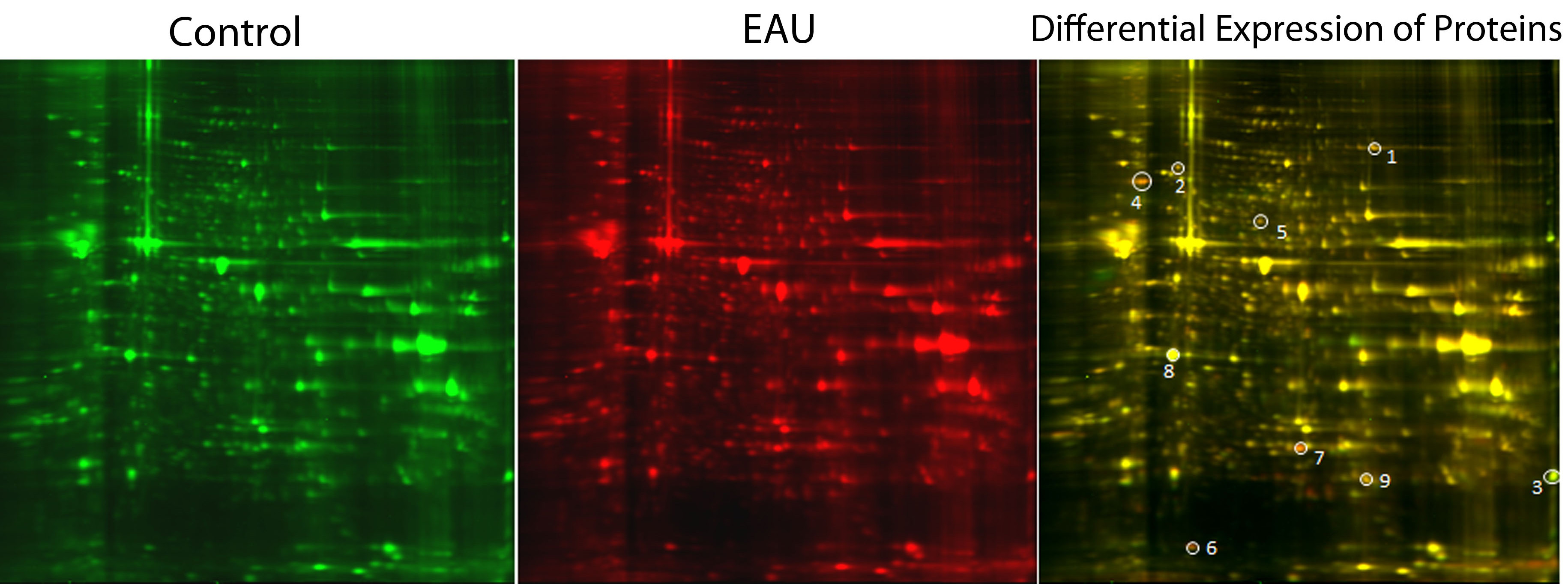
 Figure 2 of Saraswathy, Mol Vis 2011; 17:1814-1821.