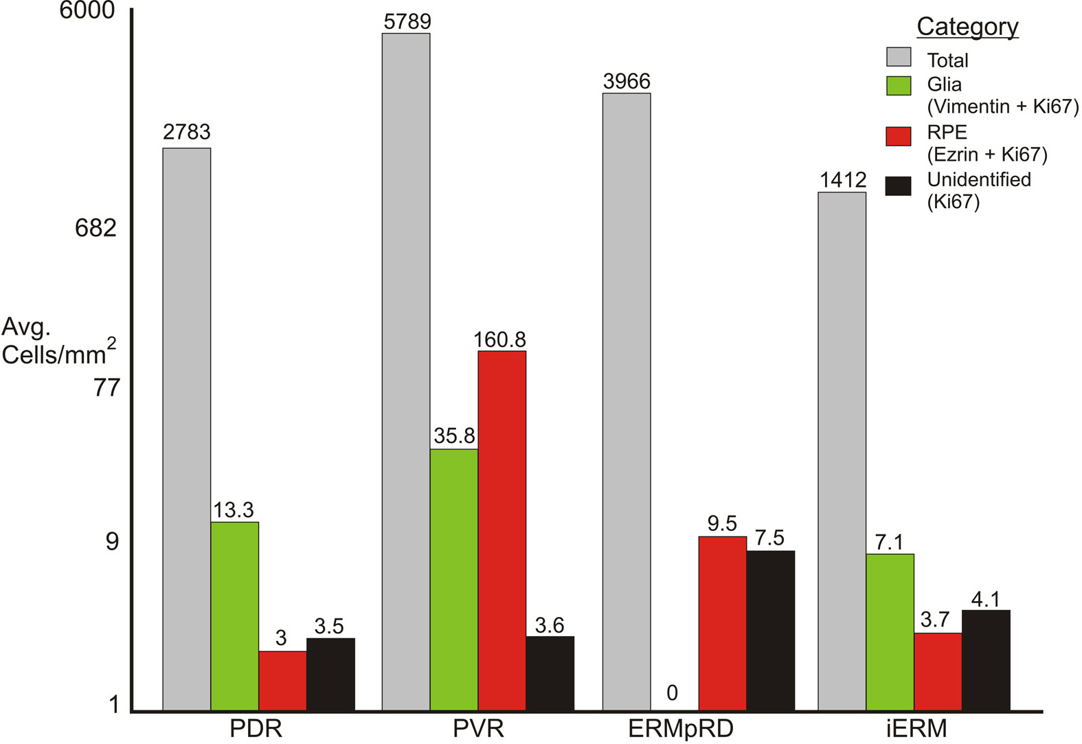
Figure 6. Illustration showing the average
number of cells/mm
2 for all four types of epiretinal
membranes. The brackets at the left of
Table 1 show which membranes
were used to generate this figure. For each membrane type (i.e.,
disease condition), the average number of cells/mm
2 equals
the total number of nuclei /mm (gray bars) divided by the number of
epiretinal membranes (ERMs) in the group. The average number of glial
cells (green bars) equals the number of vimentin-positive cells that
were also labeled with K
i-67/mm
2 in each ERM
divided by the number of ERMs in the group. The value for retinal
pigment epithelium (RPE) cells (ezrin labeled cells, red bars) was
calculated the same way, while “unidentified” equals the value for
cells that were K
i-67 positive but not labeled with any
other markers (black bars). Abbreviations: proliferative diabetic
retinopathy (PDR); proliferative vitreoretinopathy (PVR); post–retinal
detachment (ERMpRD); idiopathic ERM (iERM).
 Figure 6 of Oberstein, Mol Vis 2011; 17:1794-1805.