Figure 3. Images of representative
staining patterns on three different types of epiretinal membranes. The
membranes were from post-retinal detachment (pRD; A), of
idiopathic origin (iERM; B), and proliferative diabetic
retinopathy (PDR; C). Each membrane is labeled with anti-MIB-1
(red), anti-glial fibrillary acidic protein (GFAP; green) and ricin
(blue). In the lower half of each image the green and blue channels are
turned off to more easily see the anti-MIB-1 staining (arrows are for
reference points). Scale bars equal 50 µm.
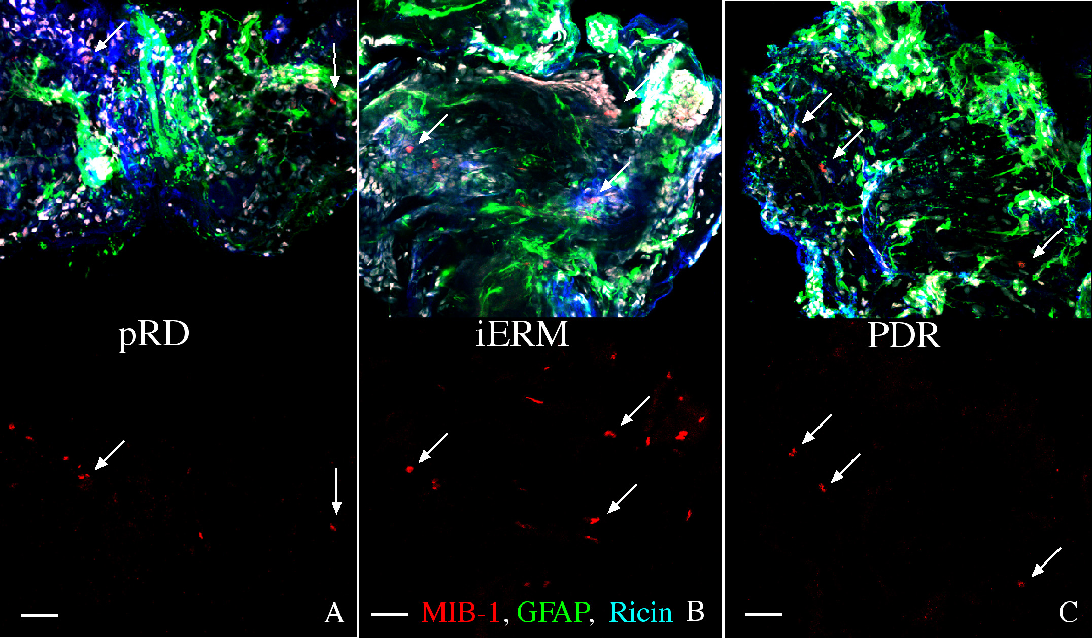
 Figure 3 of Oberstein, Mol Vis 2011; 17:1794-1805.