Figure 2. Images of representative
staining patterns on epiretinal membranes from proliferative
vitreoretinopathy. Anti-MIB-1 (A, B, F; red) or
anti-SP6 (C, D, E; red) labeling was observed
among all cell types: glia (A, B, F; green),
immune cells (A-F; blue) retinal pigment epithelial (RPE)
cells (C, D, E; green). Note that the amount of
anti-glial fibrillary acidic protein (GFAP) labeled glia varied between
membranes (A, B, F). The anti-ezrin labeling
appeared to encircle the cells, and the ricin labeling was prevalent in
all samples. Scale bars equal 50 µm.
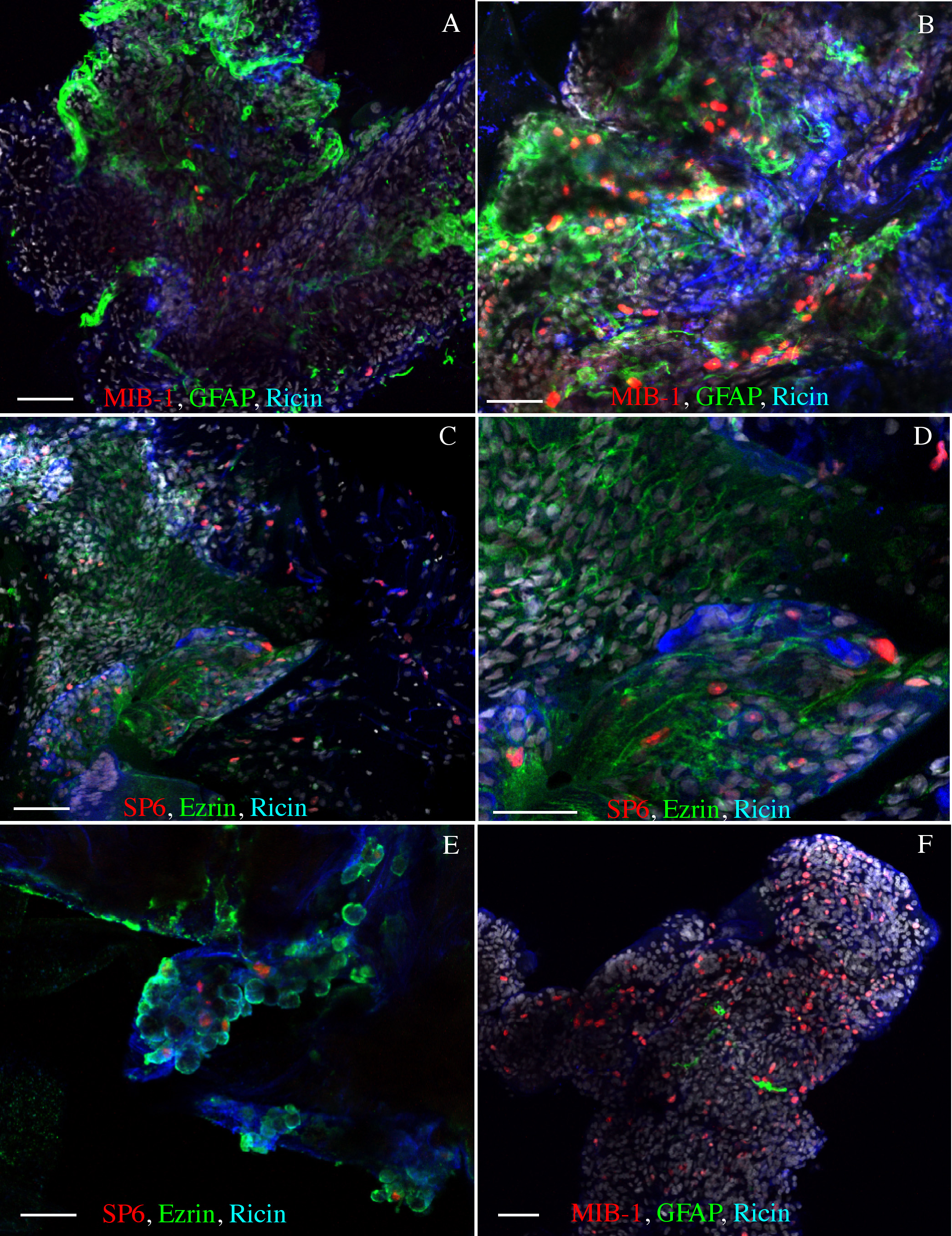
 Figure 2 of Oberstein, Mol Vis 2011; 17:1794-1805.