Figure 4. Sample confocal slices of retinas treated with AAV2.CBA.eGFP and heparinase III or chondroitin ABC lyase. GFP fluorescence
and DAPI staining are merged and the images correspond to A and D: the inner retina, B and E: the mid retina, and C and F: the outer retina. Image sizes 238×238 µm.
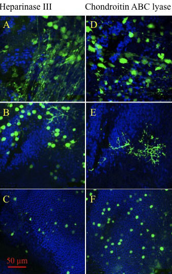
 Figure 4 of
Cehajic-Kapetanovic, Mol Vis 2011; 17:1771-1783.
Figure 4 of
Cehajic-Kapetanovic, Mol Vis 2011; 17:1771-1783.