Figure 6. Integration of murine retinal
progenior cells (mRPCs) after transplantation with a cocktail
(Chondroitinase ABC 0.01 U/ml, DAPT 10 uM, IGF1 10 ng/ ml) into Rho−/−
mice. A: The untreated control group (Synaptophysin). B:
The
untreated control group (protein kinase alpha-PKCα). The second row
(C, D, E, F) and the third row (G,
H, I, J) were cocktail treated group. The right
panel is a merged image of the three panels to the left. Integrated
cells express the synaptophysin (arrow in D), and appear to
make synaptic contact with the bipolar cells (arrow in H).
Scale bar: 20 um (A, B); 10 um (F, J).
Abbreviations: ganglion cell layer (GCL); inner plexiform layer (IPL);
inner nuclear layer (INL); and outer nuclear layer (ONL); and
subretinal space (SRS).
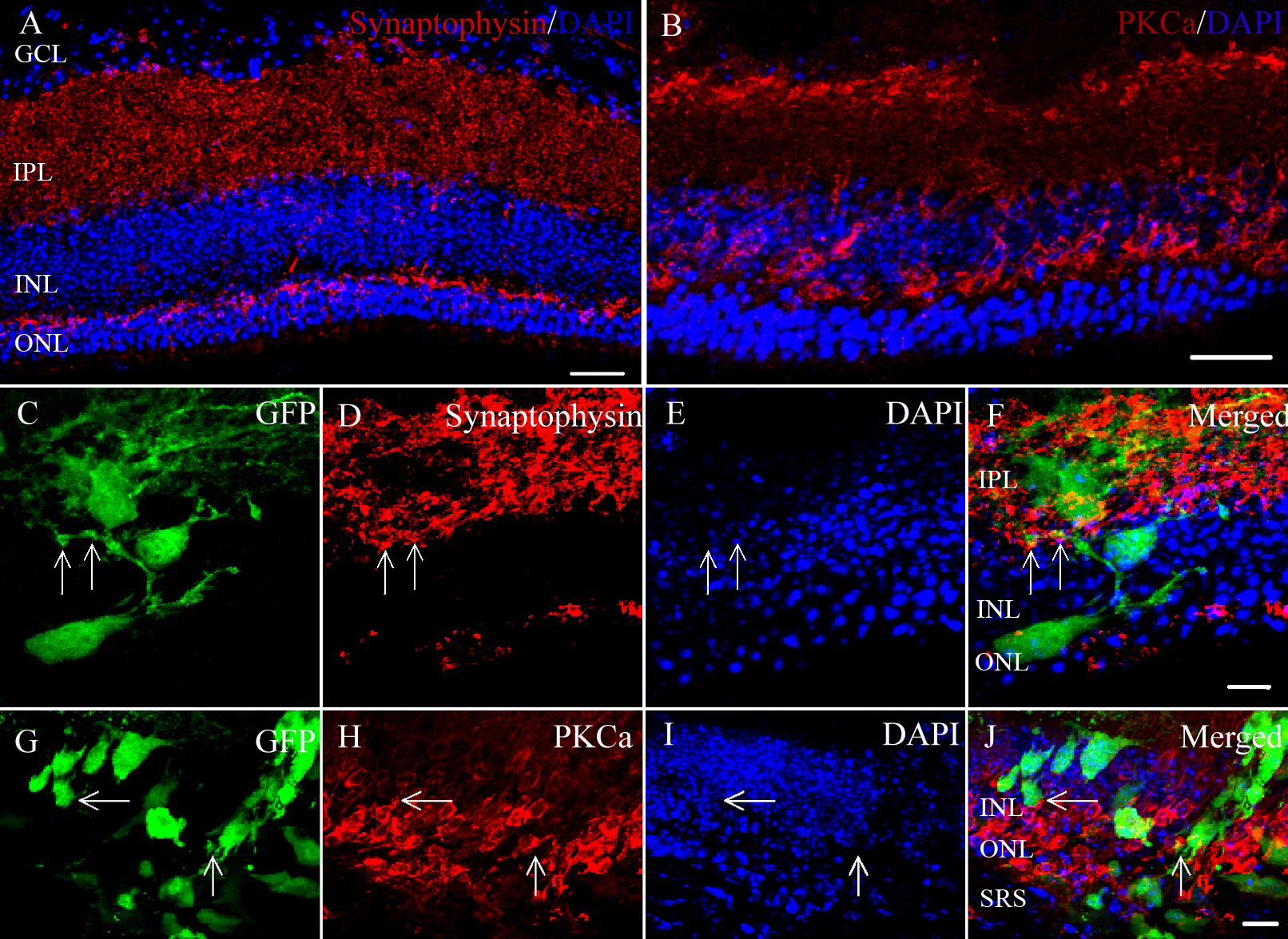
 Figure 6 of Ma, Mol Vis 2011; 17:1759-1770.