Figure 6. Retinal ganglion cell axons are
compressed by displaced retinal vessels Representative retinal
cross-sections from a control (A-C) and a photoexposed animal
processed 12 months after light exposure (ALE; D-F) doubly
immunoreacted to detect retinal vessels (red signal, A and D)
and
neurofilaments (green signal, B and E). In C
and F are shown the corresponding coupled images and
4’,6-diamidino-2-phenylindole (DAPI) counterstaining. In control
retinas, DAPI staining shows the typical layered structure of the
retina, where retinal ganglion cell (RGC) axons run parallel to the
retinal layers (B), above the RGC nuclei (C). ALE,
however, the outer nuclear layer (ONL) has disappeared, DAPI positive
nuclei are observed crossing vertically the inner plexiform layer (IPL;
F) and the RGC axons are interrupted and dragged down (E,
arrow) by retinal vessels vertically crossing the retina (D,
arrowhead). The scale bar represents 100 µm.
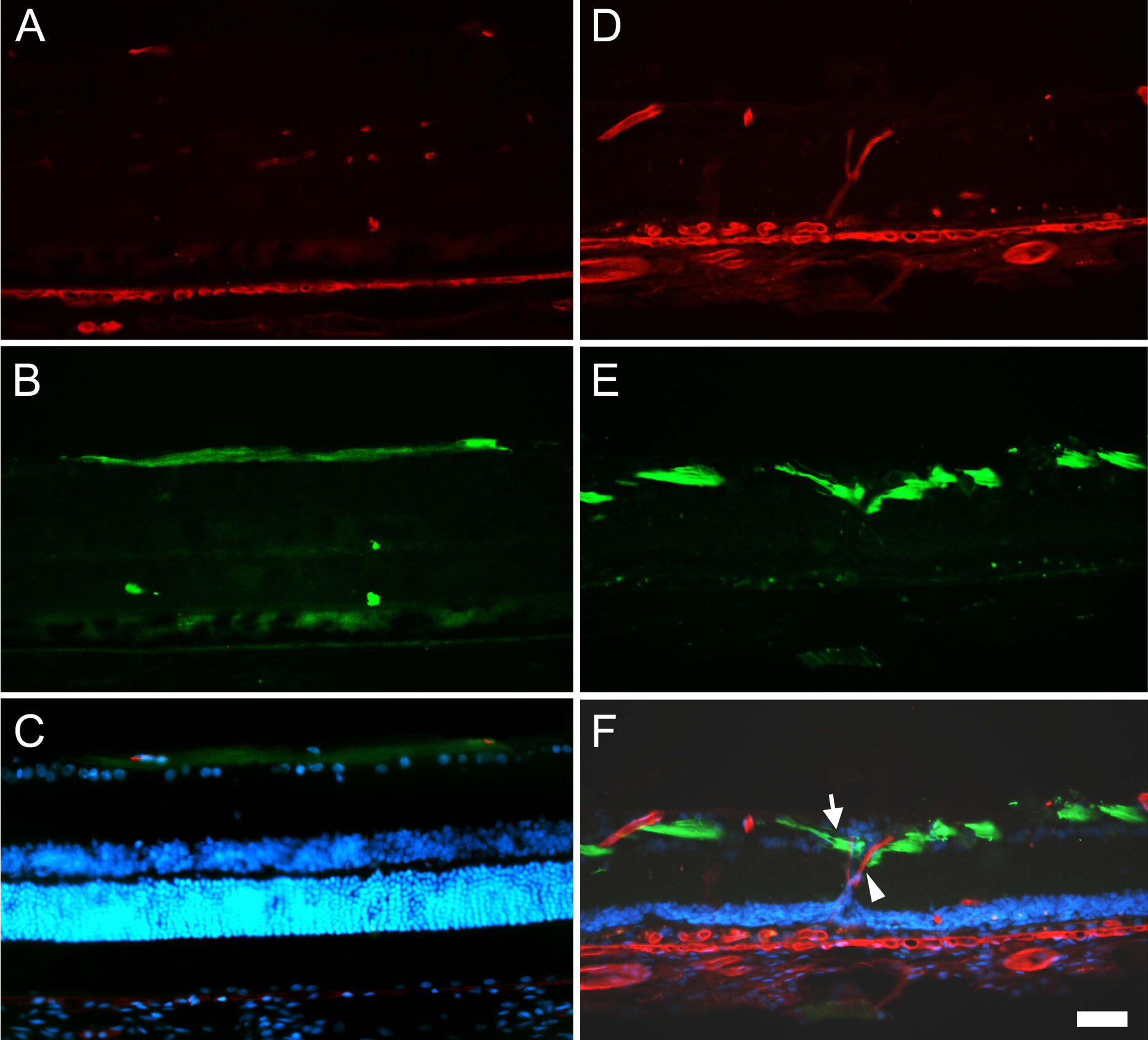
 Figure 6 of Garcia-Ayuso, Mol Vis 2011; 17:1716-1733.