Figure 5. Retinal ganglion cell axonal
abnormalities after light exposure. Photomontages of representative
phosphorylated high molecular weight neurofilament subunit (pNFH)
immunostained retinas from one control (A) and three
light-exposed animals (B-D) processed 3 (B), 9 (C),
and
12 (D) months after light exposure (ALE). The linear
trajectory of the retinal ganglion cell (RGC) axons observed in the
control retinas (A) becomes irregular in the retinas processed 3
or more (B-D) months ALE. These axonal abnormalities are
more important and severe as the time ALE increases (compare B
to D). E-J: Microphotographs of the optic nerve
fiber layer in whole mount preparations of the retinas of one control
animal (E) and five experimental animals processed 9 (F, G,
H) or 12 (I, J) months ALE. In the experimental
animals, axonal compressions by retinal vessels (F, H-J),
axonal
bulbs and meandering axons (arrowheads, H-J) and pNFH+
RGC somas (arrows, F-H) are observed. The scale bar represents
50 µm.
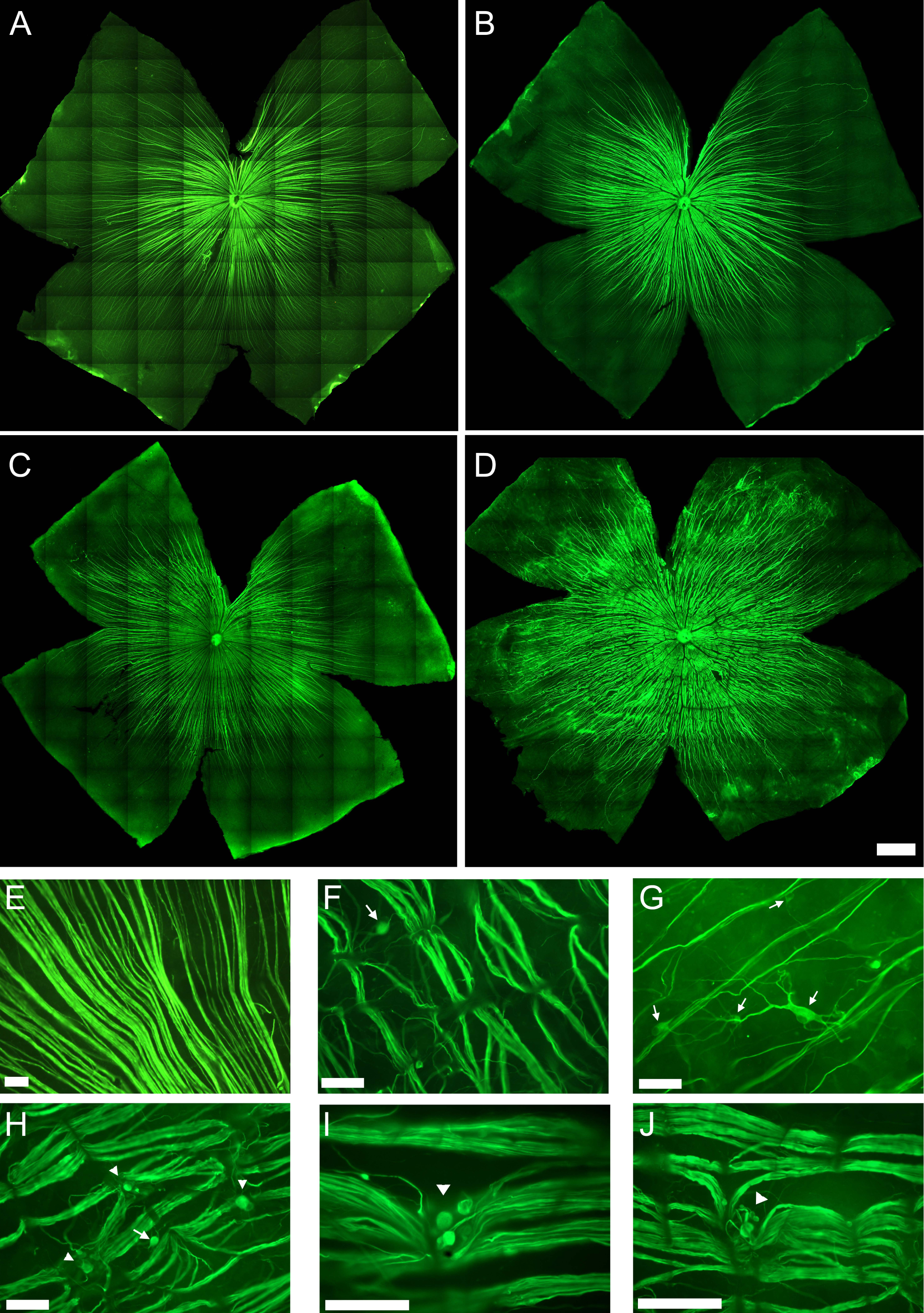
 Figure 5 of Garcia-Ayuso, Mol Vis 2011; 17:1716-1733.