Figure 6. Detection of protein expression in ARPE-19 cells after dimethyl sulfoxide or fenretinide treatment by western blot analysis.
Cells were treated with 3 μM fenretinide (FR) or dimethly sulfoxide (DMSO) for 7 days and extracts prepared for western blot.
Equal amounts of protein were loaded onto a sodium dodecyl sulfate PAGE gel, transferred to membrane, and probed with antibodies
generated against cone rod homeobox (CRX), opsin 3 (OPN3), melanopsin (OPN4), calbindin 2 (CALB2), cytokeratin 8 (KRT8), paired
box 6 (PAX6), and sex determining region Y-box 2 (SOX2). Proteins were detected using chemiluminescence and visualized using
autoradiographic film. Membranes were stripped and reprobed with glyceraldehyde 3-phosphate dehydrogenase (GAPDH) as a loading
control. Representative blots from DMSO- and FR- treated cells are shown. Protein levels were quantified using densitometry
and normalized relative to the GAPDH loading control. Data shown are mean±standard error of the mean normalized to GAPDH (n=4).
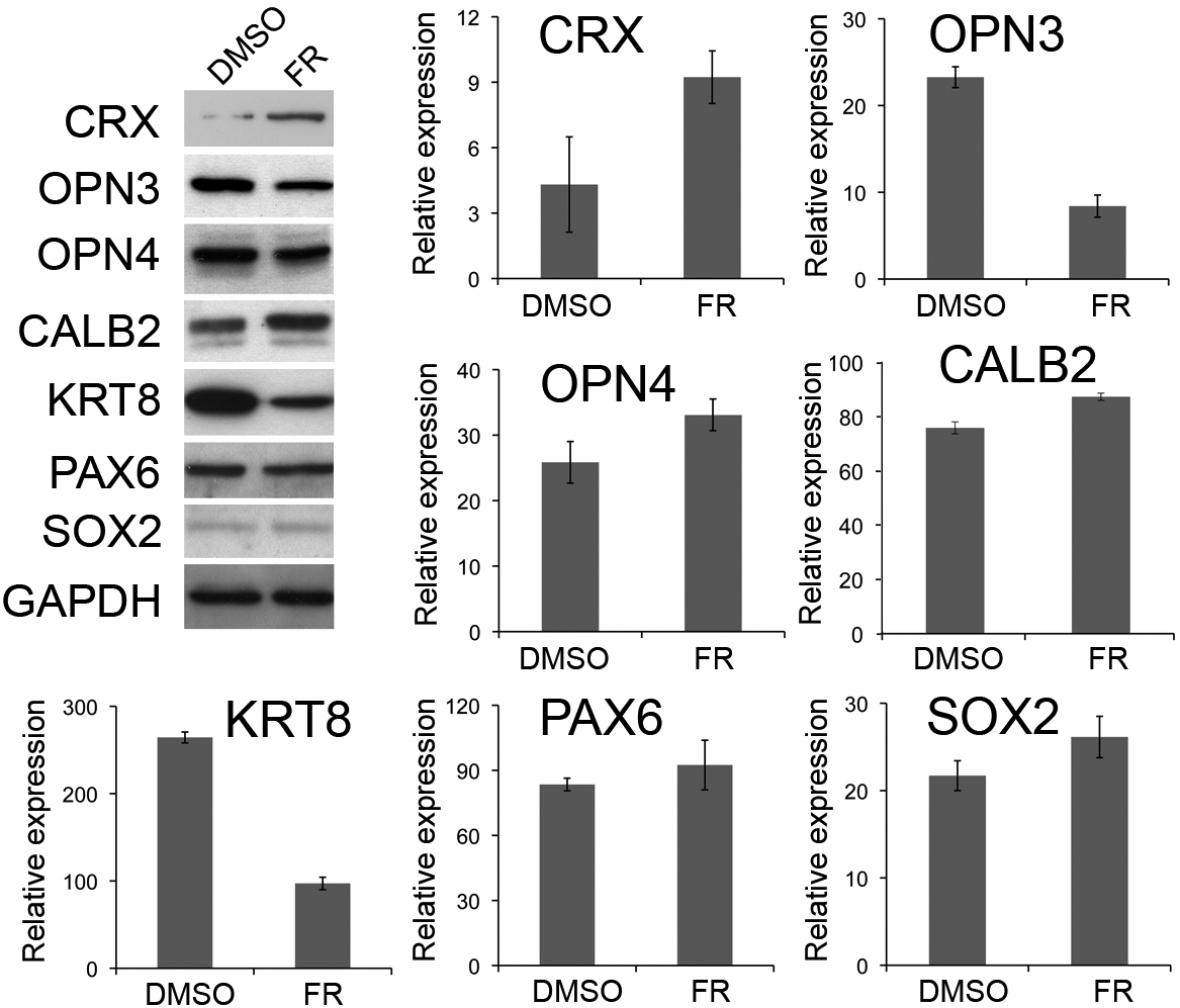
 Figure 6 of
Carr, Mol Vis 2011; 17:1701-1715.
Figure 6 of
Carr, Mol Vis 2011; 17:1701-1715.