Figure 5. Neuronal markers are expressed
in retinal pigment epithelium cells in culture but are lost after
culture on a porcine lens capsule membrane (PLC). The expression of
neurofilament heavy and medium polypeptides (NF-H and NF-M
respectively) varied in dimethyl sulfoxide (DMSO)-treated cells. A:
The
first two panels show the variation of NF-H expression within the
same dish of DMSO-treated ARPE-19 cells, which is increased across the
whole dish after fenretinide (FR) treatment. Similarly, the expression
of NF-M varies within cells of the same field and is increased by
fenretinide. B: Passage 2 primary human RPE cells and an
additional human RPE cell line (h1RPE7) express the neuronal markers
NF-H and synaptophysin (SYP). C: Culturing ARPE-19 on a PLC
decreases the expression of neuronal markers in control cells and
prevents the fenretinide-induced increase of neuronal markers. Protein
staining is indicated by the color of the text (red or green). All
scale bars equal 50 μm.
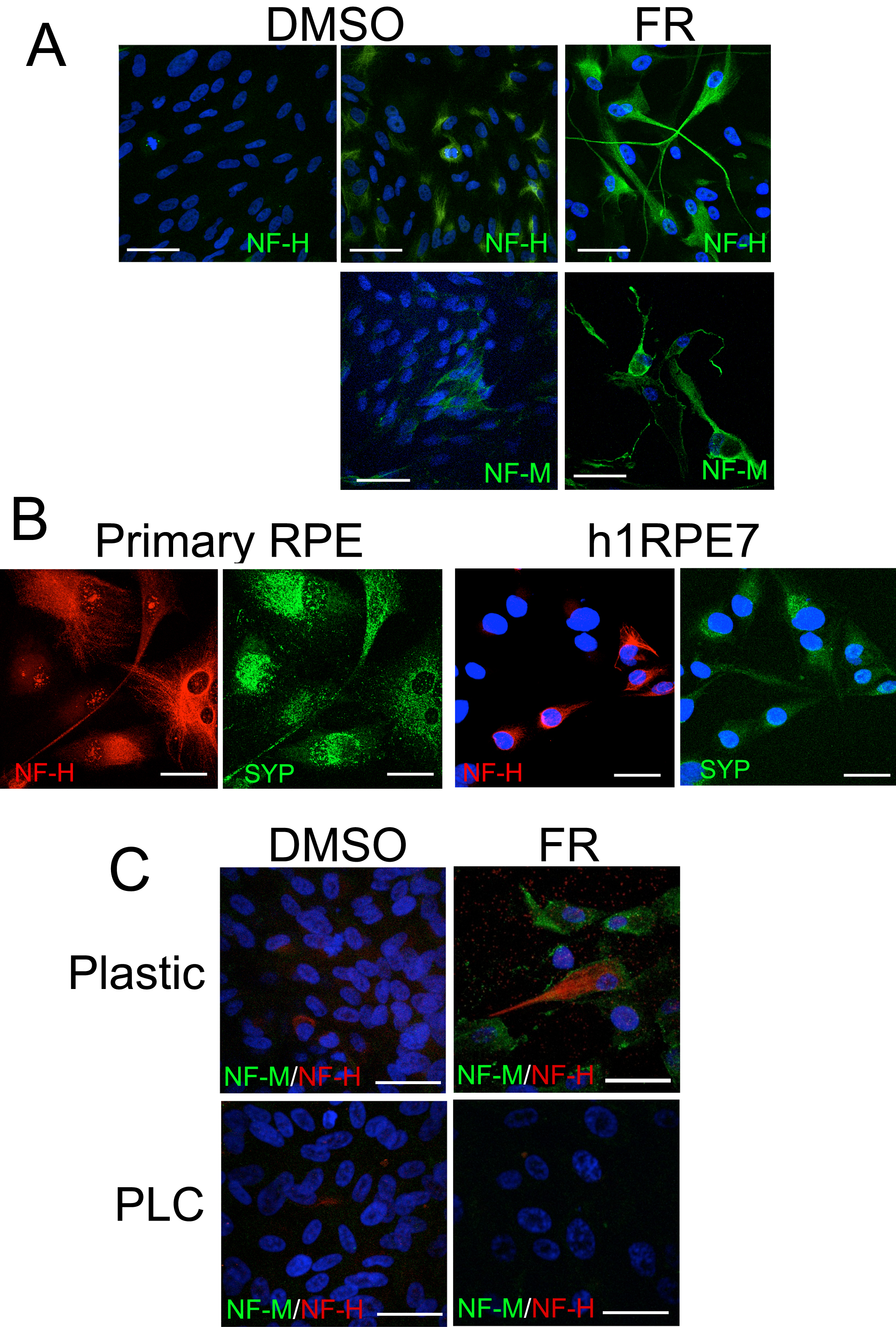
 Figure 5 of Carr, Mol Vis 2011; 17:1701-1715.