Figure 4. Immunocytochemical analysis of
neuronal and photoreceptor cell markers in ARPE-19 cells after
treatment with dimethyl sulfoxide or fenretinide. A-K: ARPE-19
cells were treated with 3 μM fenretinide or DMSO and processed for
immunocytochemistry. The nuclei of cells are stained with
4',6-diamidino-2-phenylindole (blue) with the exception of cells where
protein expression was detected within the nucleus (OPN4, CRX, and
PAX6). Staining is indicated by the text color. L: Staining was
not detected in control plates where only secondary antibodies were
used. M: Final cell density did not affect cell morphology or
changes in protein expression. ARPE-19 cells were seeded at various
densities (i, 0.5×10³; ii, 1×10³; and iii, 2×10³
cells/dish) and cultured for 7 days in DMSO-containing medium before
staining for CALB2 (red). All scale bars are 50 μm.
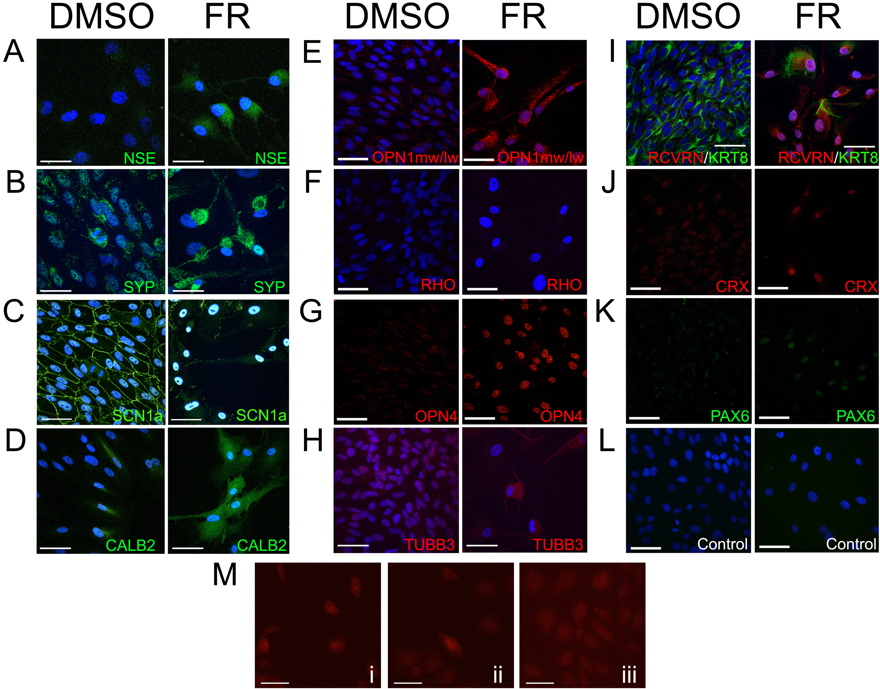
 Figure 4 of Carr, Mol Vis 2011; 17:1701-1715.