Figure 2. Anti-LYVE-1 and anti-CD31 immunochemistry. A: Immunochemistry with CD31 as the first antibody. All vessels show brown endothelium (red and green arrow). B: Immunochemistry with LYVE-1 as the first antibody; two vessels were the counterpart of the two in the left figure, one shows
a brown stained lymphatic vessel (green arrow), and the other one was inferred to be a blood vessel (red arrow). C: Normal human cornea was absent of blood vessels and lymphatic vessels.
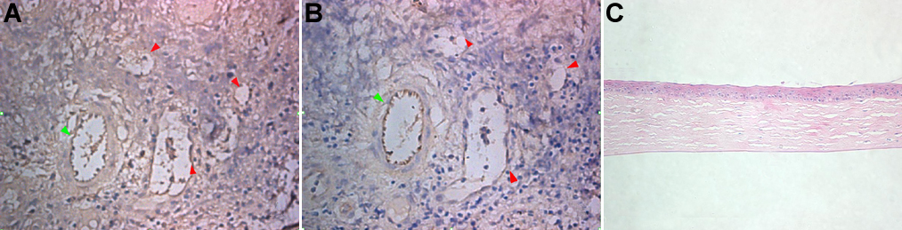
 Figure 2 of
Zheng, Mol Vis 2011; 17:1694-1700.
Figure 2 of
Zheng, Mol Vis 2011; 17:1694-1700.