Figure 2. Immunohistochemical staining for
Bcl-2, Bax, mutant p53, and TUNEL analysis positive cells in pterygium
and normal conjunctiva samples. Human pterygium tissues (A, C,
E, G); Human normal conjunctiva tissues (B, D,
F, H). Bcl-2 showed strong expression in the cytoplasm
throughout the entire width of the epithelial layer in pterygium (A).
No
expression was shown in the epithelial layer of Bcl-2 in normal
conjunctiva (B). Bax showed strong expression in the cytoplasm
throughout the entire width of the epithelial layer in both pterygium (C)
and
normal conjunctiva (D). mP53 immunostaining showed nuclear
staining in the pterygium epithelial layer (E), but no
expression in normal conjunctiva (F). For TUNEL analysis, the
positive cells in pterygium showed nuclear staining in the basal layer
of the epithelium (G). In normal conjunctiva, the positive cells
showed nuclear staining in the whole width of the epithelial layer (H).
All
slides were counterstained with Mayer’s hematoxylin. Original
magnifications: A-H, 400×.
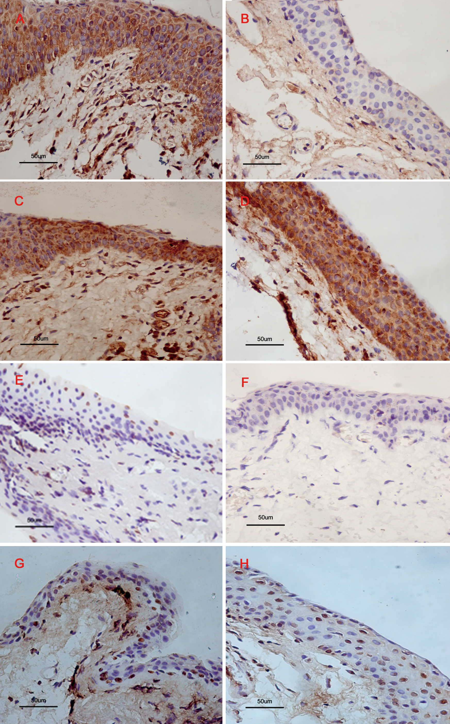
 Figure 2 of Liang, Mol Vis 2011; 17:1687-1693.