Figure 4. Versican (
VCAN) transcript analysis.
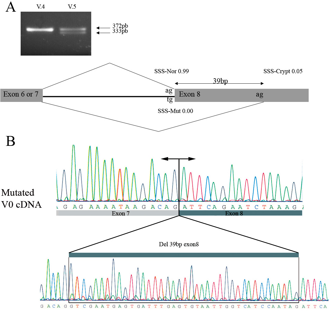
A: Ethidium bromide-stained agarose gel of reverse transcription- polymerase chain reaction (RT–PCR) products obtained with
primer pairs 7F/8R for amplification of the versican V0 mRNA from cultured lymphoblasts of one affected individual (V.5) and
one control unaffected subject (V.4). The results indicate that both a wild-type (band of 372 bp) and a 39-nt deleted (band
of 333 bp) cDNA fragment of the V0 transcript are present in the affected patient harboring the intron 7 splice acceptor mutation
of the
VCAN gene (c.4004–2A>T). The nucleotide change at the 3′ acceptor splice site of intron 7 of the
VCAN gene was analyzed using the
NNSplice program for prediction in alteration of splicing junctions, and the splice-site scores (SSSs) for a normal site (SSS-nor), a mutated
site (SSS-mut), and a cryptic site (SSS-crypt) were calculated [
28].
B: Partial sequence chromatogram of this aberrant V0 transcript lacking the first 39 bp of
VCAN exon 8.
 Figure 4 of
Brézin, Mol Vis 2011; 17:1669-1678.
Figure 4 of
Brézin, Mol Vis 2011; 17:1669-1678.