Figure 2. Mosaic multifield fundus photographs of different family members. A: Fundus photograph of patient V.2 showing a nasal dragging of the retinal vessels and fovea with white perivascular sheathing.
Clumps of pigment are visible in the nasal retina. No avascular vitreous membrane was seen. B: Fundus photograph of the right eye of patient V.6 showing a normal retinal vasculature with atrophy and pigment clumping
along and anterior to the temporal arcades. No avascular vitreous membrane was observed. C: Fundus photograph of the left eye of patient V.6 showing diffuse pigmentation with chorioretinal atrophy.
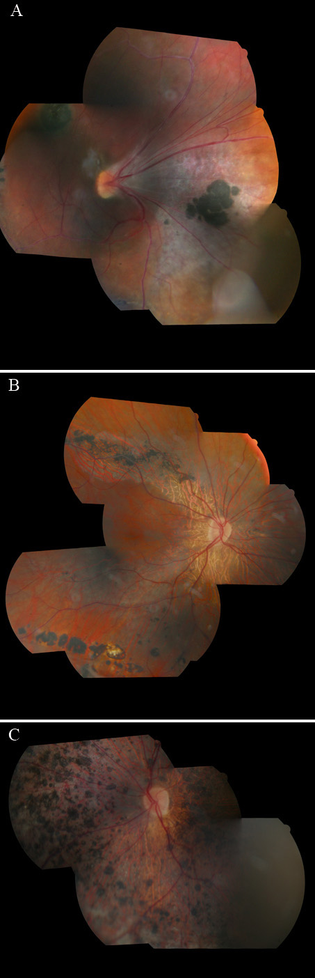
 Figure 2 of
Brézin, Mol Vis 2011; 17:1669-1678.
Figure 2 of
Brézin, Mol Vis 2011; 17:1669-1678.