Figure 4. Patterns of K 12, 13, and 19
expression in the impression cytology (IC) specimens taken from normal
sclerocorneal tissues. A, C, E, G, and H
were from corneal IC specimens. B, D, and F
were conjunctival IC specimens. Expression of K12 was present in the
corneal (A) but not in the conjunctival epithelium (B).
K13 expression was not detected in corneal epithelium (C) but
was highly expressed in the conjunctival (D) epithelium. K19 was
detected in the both corneal (E) and conjunctival (F)
epithelia. Double staining of K12/K13 (G) and K12/K19 (H)
in corneal IC specimens. Arrows: K12+/K19+ cells.
Magnification bar represents 5 μm.
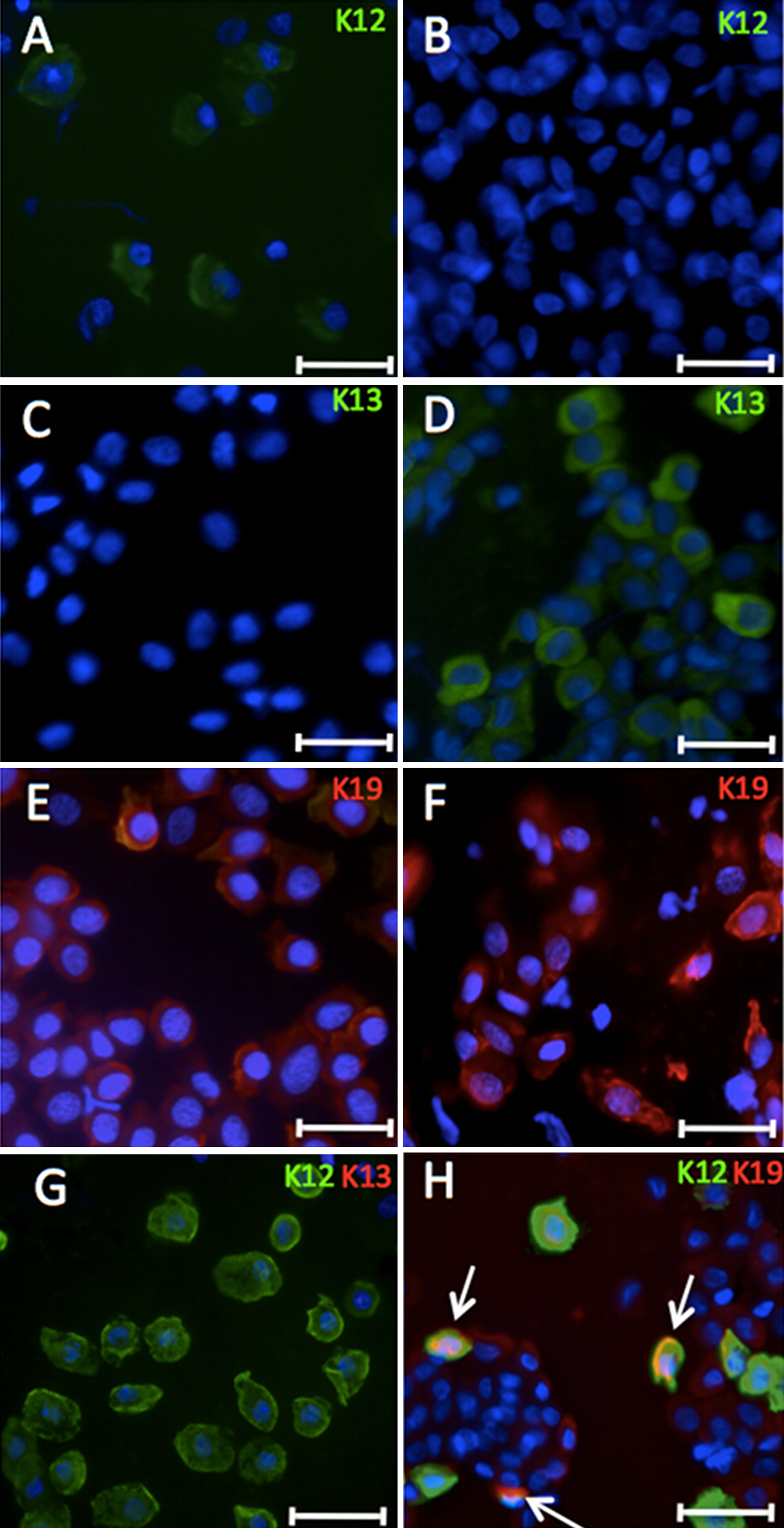
 Figure 4 of Ramirez-Miranda, Mol Vis 2011; 17:1652-1661.