Figure 2. Immunohistochemical analysis of
expression patterns of K12, K13, and K19 in normal human histologic
sections. Montage images of serial sections of the central cornea to
the conjunctiva (A-C): Double staining of K12 (green) and
K13 (red) showed the presence of K13 in the epithelia of the posterior
limbus and conjunctiva (A). K19 expression was detected in the
peripheral cornea, limbus, and conjunctiva (B). Double staining
of K12 (green) and K19 (red) showed the overlapping expression of both
cytokeratins (C). D: Expression of K12 (arrows) and K13
(arrowheads) were mutually exclusive. E: Details of the
overlapping expression of K12 and K19. Arrowhead: cells that expressed
both K12 and K19. Arrow: K12-expressing cell. Abbreviations: Conj.,
conjunctiva. Magnification bar in A, B, C
represents 200 μm. Magnification bar in D and E
represents 50 μm.
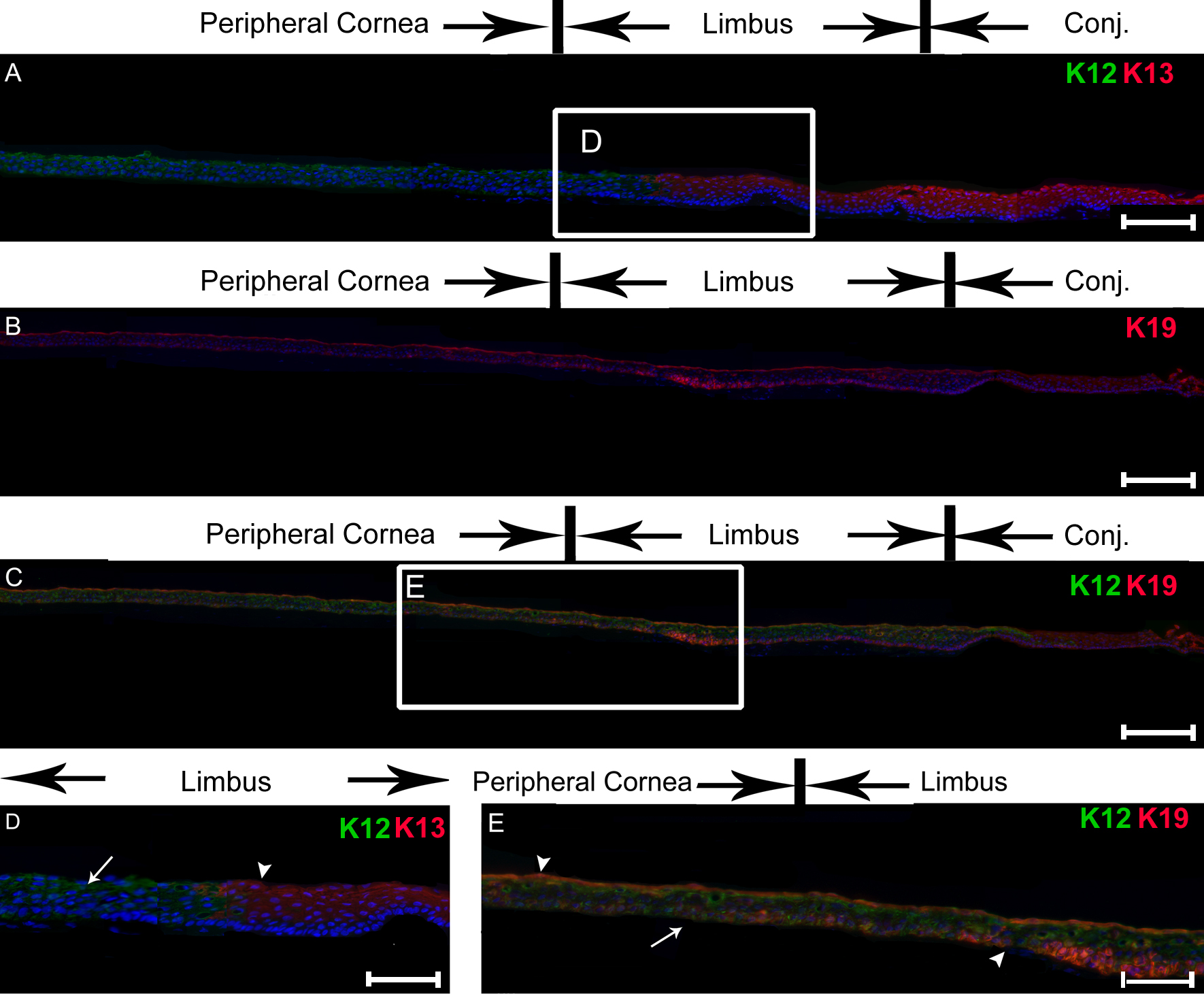
 Figure 2 of Ramirez-Miranda, Mol Vis 2011; 17:1652-1661.