Figure 6 of
Piñeiro-Gallego, Mol Vis 2011; 17:1607-1617.
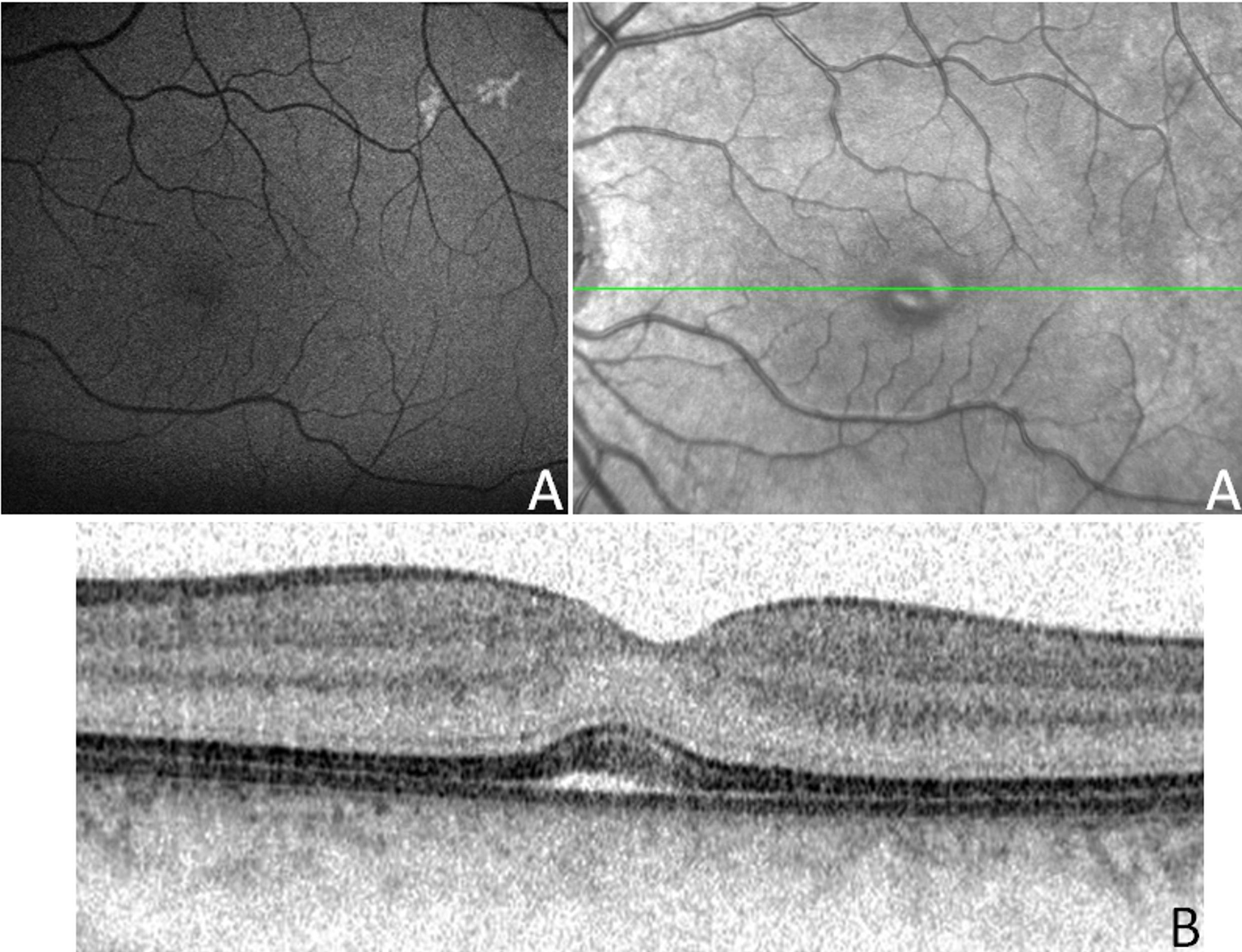
Figure 6.
Clinical evaluation of patient III:8 from the Danish family. Fundus appearance (
A
) and optical coherence tomography (OCT) showing a tiny vitelliform foveal lesion (
B
).
 Figure 6 of Piñeiro-Gallego, Mol Vis 2011; 17:1607-1617. Figure 6 of Piñeiro-Gallego, Mol Vis 2011; 17:1607-1617.