Figure 5 of
Piñeiro-Gallego, Mol Vis 2011; 17:1607-1617.
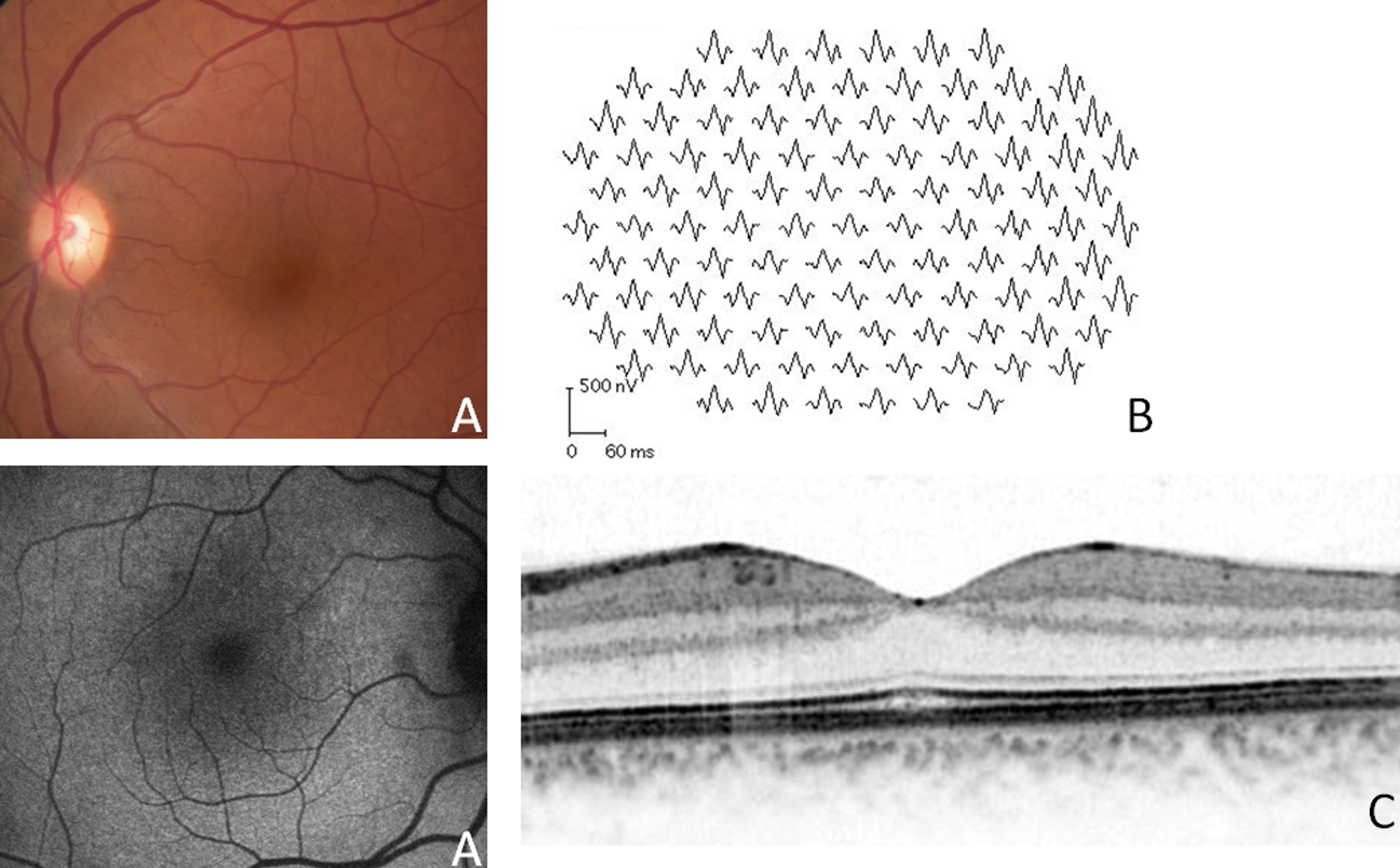
Figure 5.
Clinical evaluation of patient II:1 from the Danish family. Patient II:1 showed normal fundus appearance (
A
), multifocal-electroretinograms (ERGs;
B
), and optical coherence tomography (OCT;
C
).
 Figure 5 of Piñeiro-Gallego, Mol Vis 2011; 17:1607-1617. Figure 5 of Piñeiro-Gallego, Mol Vis 2011; 17:1607-1617.