Figure 3. Clinical evaluation of the male
sibling from the Spanish family. Color fundus appearance photographs
showing perimacular scars in both eyes (A, B). By
fluorescein angiography (AGF), there are multiple hypofluorescent discs
in the peripheral retina caused by the deposition of vitelliform
material. AGF also shows a hyperfluorescence window defect due to
atrophy of the retinal pigment epithelium (RPE). Multiple
hypoautofluorescent lesions in the peripheral retina due to lipofuscin
deposits on the fundus autofluoresence image (E, F).
Optical coherence tomography (OCT) showing RPE detachment and cystic
degeneration of the sensory retina, typical of advanced stages of
retinal dystrophies (G, H). OD: Right eye, OS: Left eye.
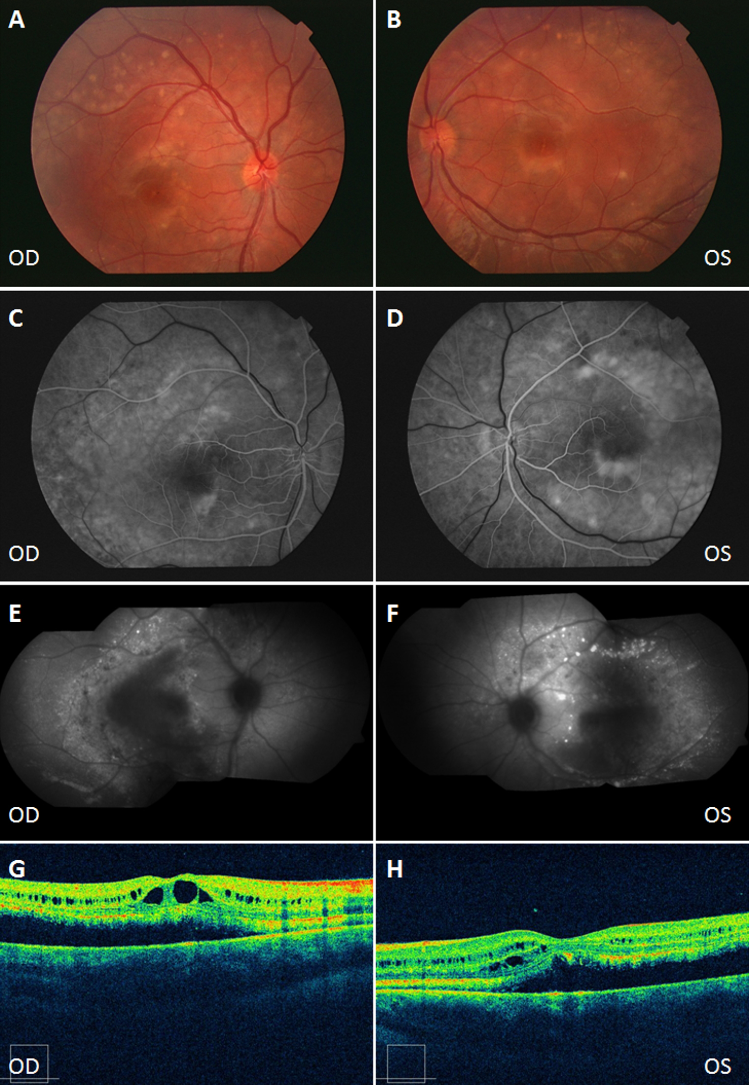
 Figure 3 of Piñeiro-Gallego, Mol Vis 2011; 17:1607-1617.