Figure 2. Clinical evaluation of the
female sibling from the Spanish family. Color fundus appearance
photographs (A, B). The right eye shows a typical
pseudohypopyon stage with a deposit of yellowish vitelliform material
in the lower half of the macular lesion. The left eye shows a
characteristic atrophic stage. Fluorescein angiography (AGF) shows a
hyperfluorescence window defect due to atrophy of the retinal pigment
epithelium (RPE; C, D), and hypofluorescence due to a
deposit of lipofuscin (C). Vitelliform lesions appear as areas
of hyperautofluorescence (E) and hypoautofluorescence (F)
fundus autofluorescence (FAF). Optical coherence tomography (OCT)
section of the pseudohypopyon lesion shows a hyperreflective material
beneath the retina in the right eye (G) and a neurosensory
retinal detachment in the left eye (H). OD: Right eye, OS: Left
eye.
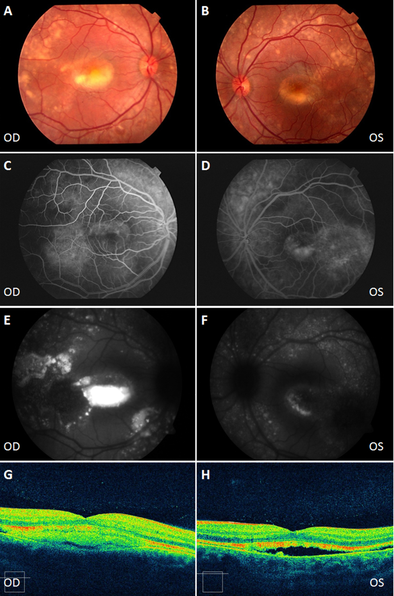
 Figure 2 of Piñeiro-Gallego, Mol Vis 2011; 17:1607-1617.