Figure 3. Ophthalmic phenotypic
characterization of the right (OD) and left (OS) eye of the index
patient. A: Color fundus photographs showing widespread changes
in the midperiphery associated with atrophic changes in the macular
area. B: Fundus autofluorescence imaging showing loss of
autofluorescence in the midperiphery and in the perifoveal region. C:
Sprectral
domain optical coherence tomography of the right (OD) and
left (OS) macula showing perifoveal thinning of the external layer of
the retina.
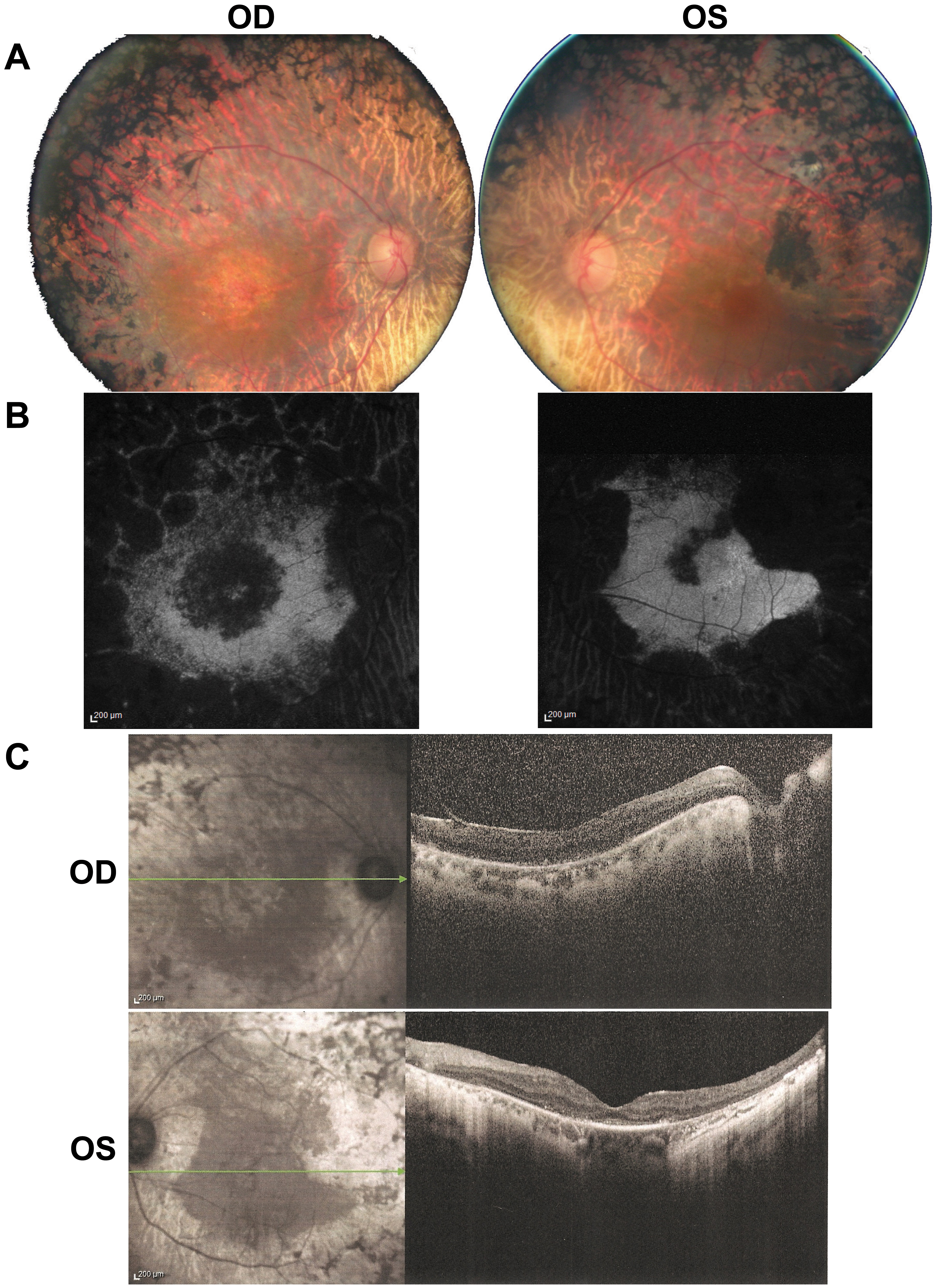
 Figure 3 of Audo, Mol Vis 2011; 17:1598-1606.