Figure 2. Tissue labeling of PEDF
mRNA and protein in rat ROP model at P14. Sections of P14 rat retina in
ROP model labeled by in situ hybridization for PEDF mRNA
showing message in the ganglion cell layer (GCL), and in the inner and
outer nuclear layers (INL, ONL; A, C). Control using
sense probe showed no signal (B, D). Immunostaining for
PEDF protein in P14 ROP model showed labeling in the inner and outer
plexiform layers and inner and outer nuclear layers (E). No
primary control showed nonspecific staining in the ganglion cell layer (F).
Magnification
calibration
bars
are 100 µm (A, B) and 50
µm (C-F).
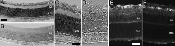
 Figure 2 of Hartmann, Mol Vis 2011; 17:1577-1587.