Figure 2. Depletion of protein kinase C
(PKC)δ prevented ethambutol (EMB)-induced vacuolar formation. Retinal
pigment epithelium (RPE)50 (A) or ARPE19 (C) and (D)
were
transfected with none (MOCK), shRNA of lamin (control shRNA), or
combinations of shRNA fragments of PKCα (90/92 in A), PKC βII (D/A2 in A
and C), or PKCδ (F/H, G/H in A and E/G in C), for 36 h followed
by treatment with 0.8 mM EMB for 24 h. Pictures were taken under a
phase contrast microscope with 200× (A, C) and 400× (D)
magnification.
(B) and (E) are quantitations of
cytoplasmic vacuolization for (A) and (D), respectively,
performed as described in Methods. These results are representative of
five reproducible experiments. The number of vacuoles/per field were
compared between indicated treatment groups using one way ANOVA
followed by Dunnett’s post hoc tests. The asterisks (* and **) indicate
statistical significance (p<0.05 and p<0.005, respectively, n=5)
between the indicated groups.
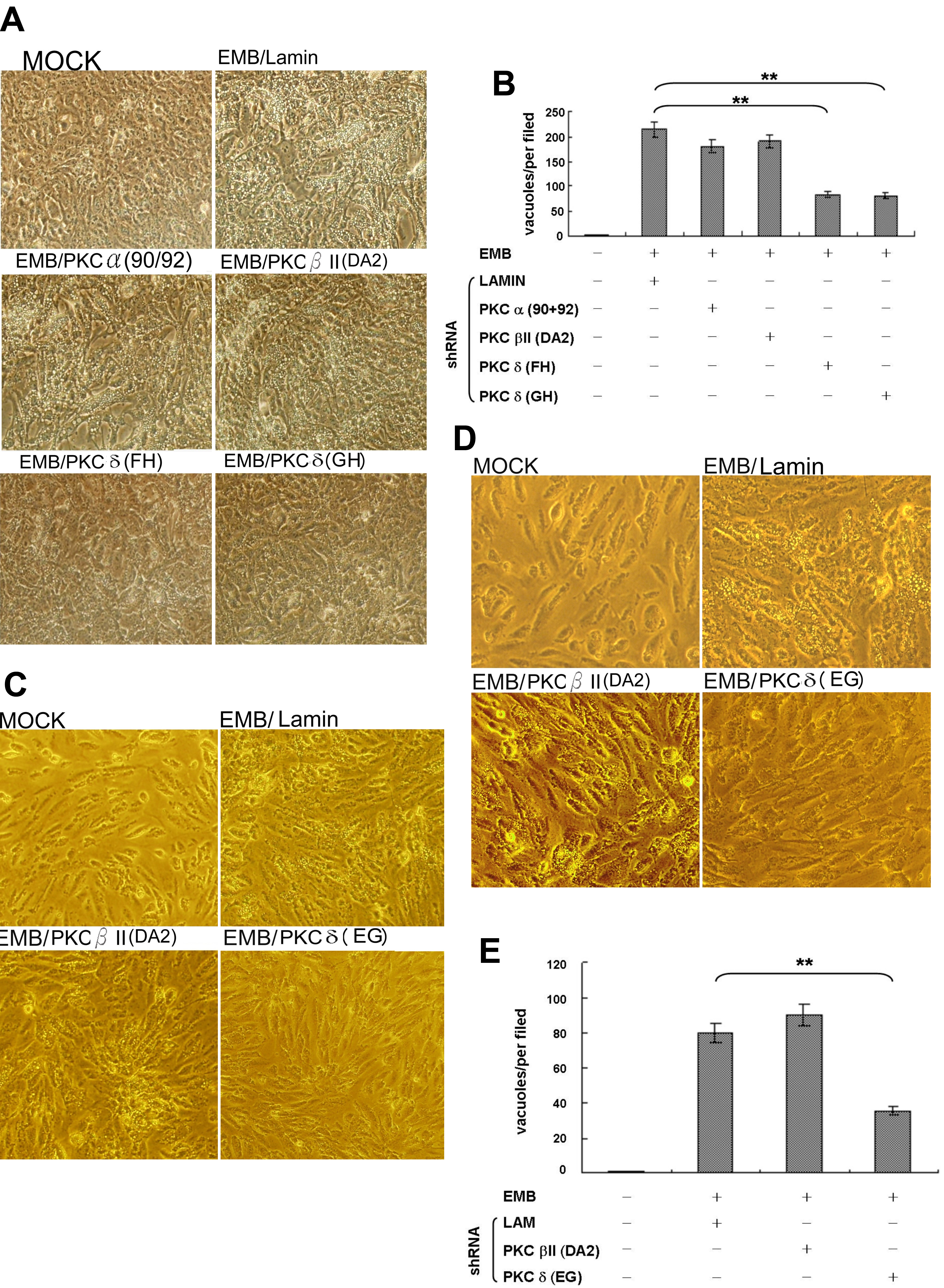
 Figure 2 of Tsai, Mol Vis 2011; 17:1564-1576.