Figure 1. Robo1 and slit2 expression in fibrovascular membranes from a 66-year-old patient with a 15-year history of diabetes. A, B, E, F: Immunofluorescent staining shows Robo1-positive (A), slit2-positive (E), and cytokeratin-positive (B, F) staining in fibrovascular membranes (FVMs). C, D, G, H: Cell nuclei were stained with 4’, 6’ -diamino-2-phenylindole (DAPI; C, G), and the colocalization of Robo1 and pancytokeratin (D), and slit2 and cytokeratin (H) are also shown. Bar graphs denote 100 μm.
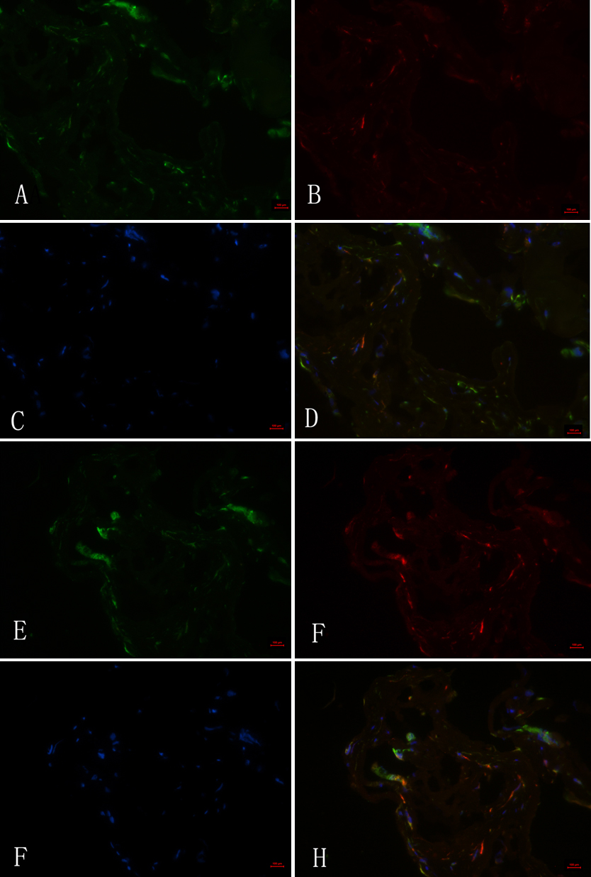
 Figure 1 of
Zhou, Mol Vis 2011; 17:1526-1536.
Figure 1 of
Zhou, Mol Vis 2011; 17:1526-1536.