Figure 3. The distribution of Dexras1 expression and the change in outer nuclear layer thickness after light exposure. Slides were examined
at 200× or 400× magnification on a Leica light microscope. Dexras1 expression and outer nuclear layer (ONL) thickness were
observed in sections of A: negative control retina (primary antibody was substituted by PBS), B: normal retina, and C: injured retina at 1 day after light exposure by immunohistochemistry. D-F: Magnified images for A-C, respectively, are represented. G: Quantitative results for numbers of Dexras1 positive cells are at a lower magnification. H: Quantitative analysis of ONL thickness between the normal retina and injured retina was conducted. Values are expressed as
mean±SEM (n=3 animals, 6 eye samples from three animals for each group, *p<0.05). Abbreviations: GCL represents ganglion cell
layer; INL represents inner nuclear layer; ONL represents outer nuclear layer.
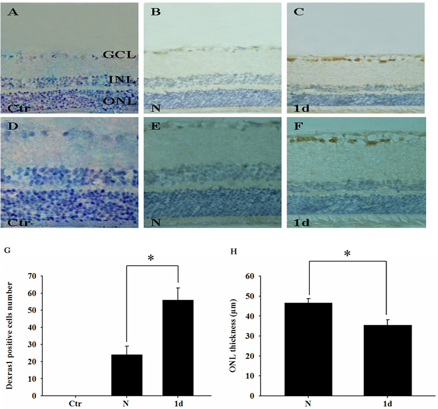
 Figure 3 of
Sang, Mol Vis 2011; 17:134-143.
Figure 3 of
Sang, Mol Vis 2011; 17:134-143.