Figure 4. Status of the RGC during EAU. Tuj-1 staining (green) by an antibody against neuronal β3-tubulin in cell bodies and axons showed
microtubule stability of the neuronal cells. DAPI (blue) was applied to counterstain the cell nuclei. A: During the course of EAU, Tuj-1-positive staining increased and spread at 7 dpi, appeared disrupted at the peak of inflammation at 14 dpi, and became the
most intensive at 21 dpi with the restoration of normal retinal architecture. The staining was then downregulated at 28 dpi
but became relatively stronger again at 35 dpi. The size bar represents 200 μm (10×). B: High magnification observation (40×) on the Tuj-1-positive staining showed that, while microtubules of the neuronal axons
were destabilized and disassembled when the disease progressed, they gradually restored their features at later stages of
EAU. Compared to the staining of the control and that at 28 dpi, distribution of Tuj-1-staining at 35 dpi indicated that the
neuronal activities underwent waves of adjustment at the later phase of disease. C: Relative expression of thymocyte differentiation antigen 1 (Thy-1) and growth-associated protein-43 (Gap-43) during EAU
by real-time RT–PCR. Data represent at least three repeated experiments. RPE, retinal pigment epithelium; ONL, outer nuclear
layer; INL, inner nuclear layer; RGC, retina ganglion cells. The size bar represents 50 μm (40×).
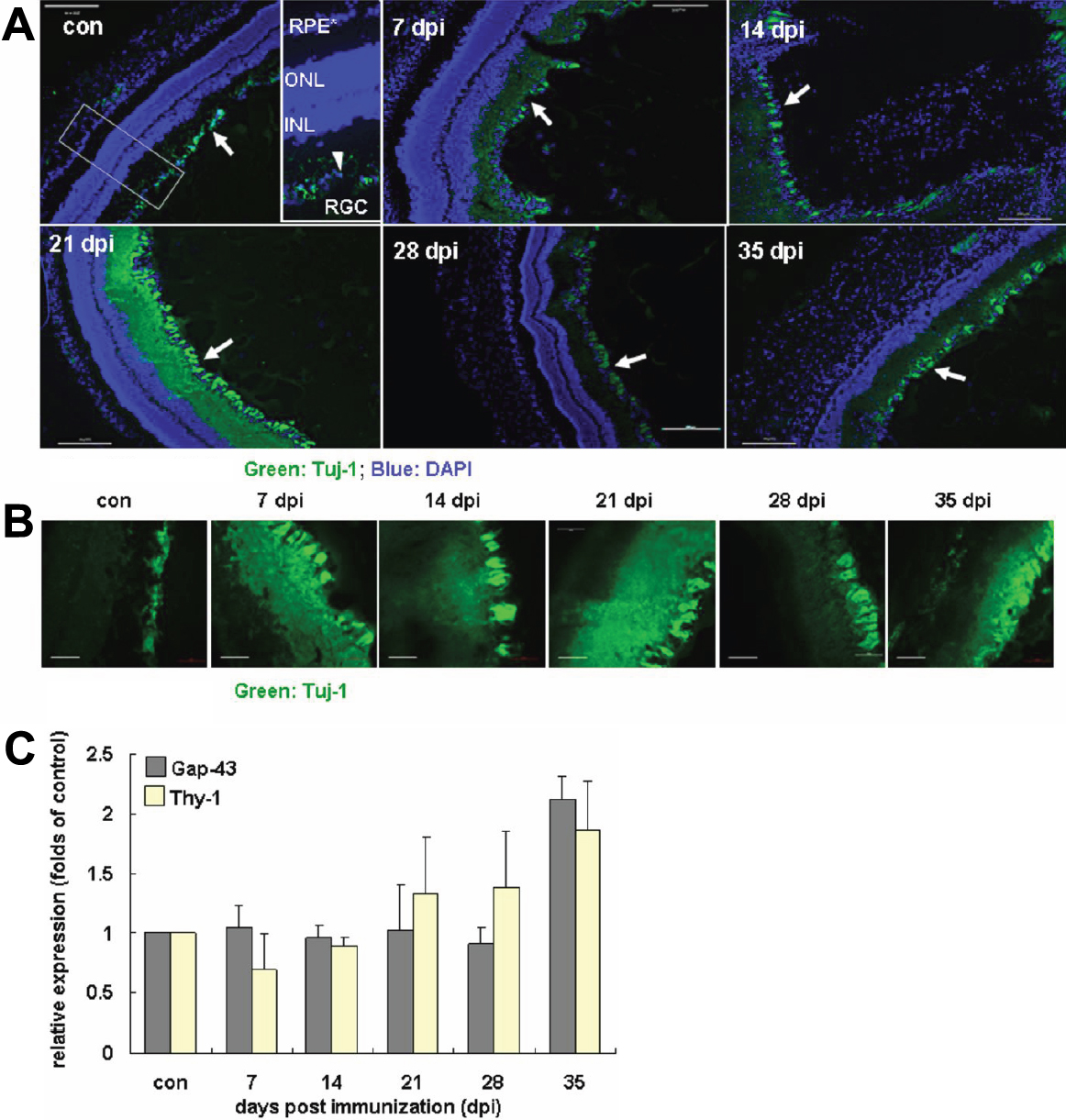
 Figure 4 of
Jia, Mol Vis 2011; 17:1493-1507.
Figure 4 of
Jia, Mol Vis 2011; 17:1493-1507.