Figure 3. Activation patterns of resident retinal astrocytes and Müller cells during EAU. The antibody against glial fibrillary acidic
protein (GFAP; green) was used to identify astrocytes and Müller cells; 2-(4-amidinophenyl)-6-indolecarbamidine dihydrochloride
(DAPI; blue) was applied to counterstain the cell nuclei. In the CFA control (A) and EAU 7 dpi group (B), there were no detectable GFAP in the endfeet of Müller cells (ILM), and GFAP staining was limited within astrocytes (NFL).
Panels C (14 dpi), D (21 dpi), E (28 dpi), and F (35 dpi) show that GFAP became detectable throughout the Müller cell cytoplasm from INL to ONL (bidirectional arrows). In
addition, positive staining widened and enlarged in astrocytes (arrows), which turned pleomorphic (hypertrophy of the cell
body and nucleus, elongation of cytoplasmic processes, irregular in shape) and continuously activated from 14 to 35 dpi. Data
represent at least three repeated experiments. OLM, outer limiting membrane; ONL, outer nuclear layer; INL, inner nuclear
layer; ILM, inner limiting membrane; NFL, nerve fiber layer. The size bar represents 200 μm (10×).
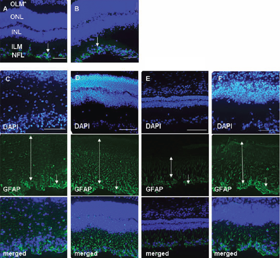
 Figure 3 of
Jia, Mol Vis 2011; 17:1493-1507.
Figure 3 of
Jia, Mol Vis 2011; 17:1493-1507.